Calcium »
PDB 2agq-2b03 »
2aik »
Calcium in PDB 2aik: Formylglycine Generating Enzyme C336S Mutant Covalently Bound to Substrate Peptide Lctpsra
Protein crystallography data
The structure of Formylglycine Generating Enzyme C336S Mutant Covalently Bound to Substrate Peptide Lctpsra, PDB code: 2aik
was solved by
D.Roeser,
M.G.Rudolph,
with X-Ray Crystallography technique. A brief refinement statistics is given in the table below:
| Resolution Low / High (Å) | 29.71 / 1.73 |
| Space group | P 21 21 2 |
| Cell size a, b, c (Å), α, β, γ (°) | 61.719, 109.517, 43.520, 90.00, 90.00, 90.00 |
| R / Rfree (%) | 14.1 / 17.4 |
Other elements in 2aik:
The structure of Formylglycine Generating Enzyme C336S Mutant Covalently Bound to Substrate Peptide Lctpsra also contains other interesting chemical elements:
| Chlorine | (Cl) | 1 atom |
Calcium Binding Sites:
The binding sites of Calcium atom in the Formylglycine Generating Enzyme C336S Mutant Covalently Bound to Substrate Peptide Lctpsra
(pdb code 2aik). This binding sites where shown within
5.0 Angstroms radius around Calcium atom.
In total 2 binding sites of Calcium where determined in the Formylglycine Generating Enzyme C336S Mutant Covalently Bound to Substrate Peptide Lctpsra, PDB code: 2aik:
Jump to Calcium binding site number: 1; 2;
In total 2 binding sites of Calcium where determined in the Formylglycine Generating Enzyme C336S Mutant Covalently Bound to Substrate Peptide Lctpsra, PDB code: 2aik:
Jump to Calcium binding site number: 1; 2;
Calcium binding site 1 out of 2 in 2aik
Go back to
Calcium binding site 1 out
of 2 in the Formylglycine Generating Enzyme C336S Mutant Covalently Bound to Substrate Peptide Lctpsra
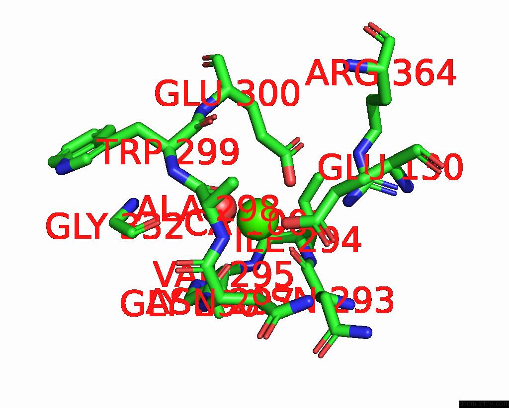
Mono view
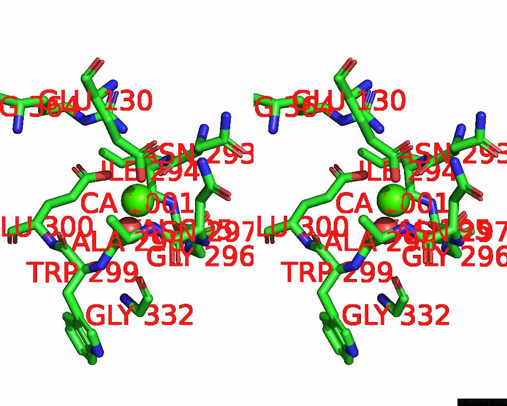
Stereo pair view
Mono view
Stereo pair view
A full contact list of Calcium with other atoms in the Ca binding
site number 1 of Formylglycine Generating Enzyme C336S Mutant Covalently Bound to Substrate Peptide Lctpsra within 5.0Å range:
|
Calcium binding site 2 out of 2 in 2aik
Go back to
Calcium binding site 2 out
of 2 in the Formylglycine Generating Enzyme C336S Mutant Covalently Bound to Substrate Peptide Lctpsra
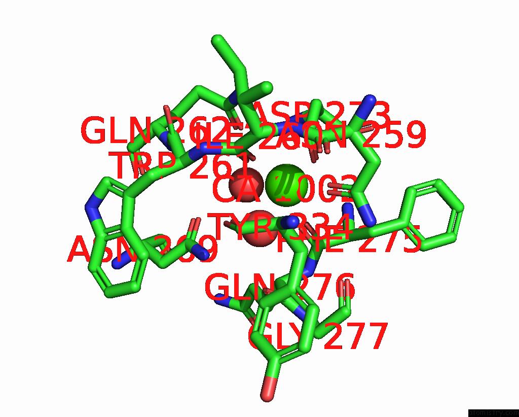
Mono view
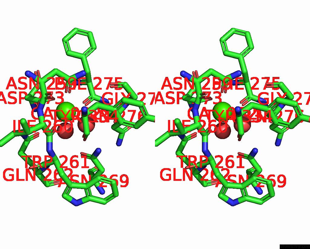
Stereo pair view
Mono view
Stereo pair view
A full contact list of Calcium with other atoms in the Ca binding
site number 2 of Formylglycine Generating Enzyme C336S Mutant Covalently Bound to Substrate Peptide Lctpsra within 5.0Å range:
|
Reference:
D.Roeser,
A.Preusser-Kunze,
B.Schmidt,
K.Gasow,
J.G.Wittmann,
T.Dierks,
K.Von Figura,
M.G.Rudolph.
A General Binding Mechanism For All Human Sulfatases By the Formylglycine-Generating Enzyme Proc.Natl.Acad.Sci.Usa V. 103 81 2006.
ISSN: ISSN 0027-8424
PubMed: 16368756
DOI: 10.1073/PNAS.0507592102
Page generated: Tue Jul 8 04:20:08 2025
ISSN: ISSN 0027-8424
PubMed: 16368756
DOI: 10.1073/PNAS.0507592102
Last articles
Ir in 3SD3Ir in 3PK2
Ir in 3F2T
Ir in 3IRW
Ir in 3D0U
Ir in 2GIS
Ir in 3B31
Ir in 1K26
Ir in 2B0U
Ir in 1C1K