Calcium »
PDB 2agq-2b03 »
2aoq »
Calcium in PDB 2aoq: Crystal Structure of Muth-Unmethylated Dna Complex
Protein crystallography data
The structure of Crystal Structure of Muth-Unmethylated Dna Complex, PDB code: 2aoq
was solved by
J.Y.Lee,
J.Chang,
N.Joseph,
R.Ghirlando,
D.N.Rao,
W.Yang,
with X-Ray Crystallography technique. A brief refinement statistics is given in the table below:
| Resolution Low / High (Å) | 19.85 / 2.20 |
| Space group | P 63 |
| Cell size a, b, c (Å), α, β, γ (°) | 114.624, 114.624, 46.755, 90.00, 90.00, 120.00 |
| R / Rfree (%) | 19.9 / 22 |
Calcium Binding Sites:
The binding sites of Calcium atom in the Crystal Structure of Muth-Unmethylated Dna Complex
(pdb code 2aoq). This binding sites where shown within
5.0 Angstroms radius around Calcium atom.
In total 2 binding sites of Calcium where determined in the Crystal Structure of Muth-Unmethylated Dna Complex, PDB code: 2aoq:
Jump to Calcium binding site number: 1; 2;
In total 2 binding sites of Calcium where determined in the Crystal Structure of Muth-Unmethylated Dna Complex, PDB code: 2aoq:
Jump to Calcium binding site number: 1; 2;
Calcium binding site 1 out of 2 in 2aoq
Go back to
Calcium binding site 1 out
of 2 in the Crystal Structure of Muth-Unmethylated Dna Complex
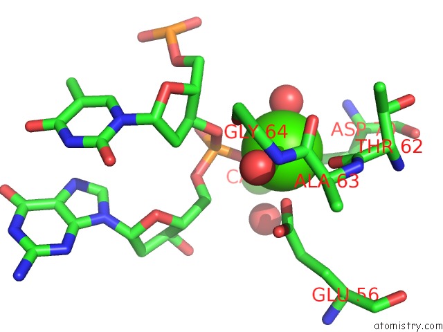
Mono view
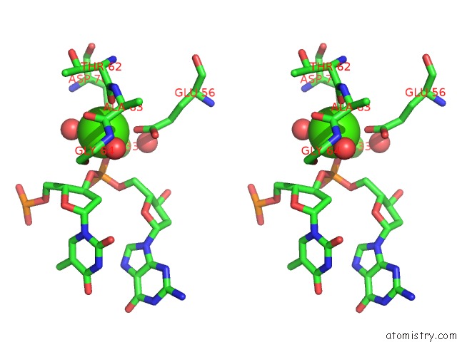
Stereo pair view
Mono view
Stereo pair view
A full contact list of Calcium with other atoms in the Ca binding
site number 1 of Crystal Structure of Muth-Unmethylated Dna Complex within 5.0Å range:
|
Calcium binding site 2 out of 2 in 2aoq
Go back to
Calcium binding site 2 out
of 2 in the Crystal Structure of Muth-Unmethylated Dna Complex
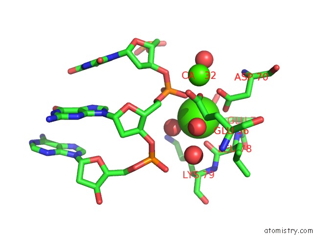
Mono view
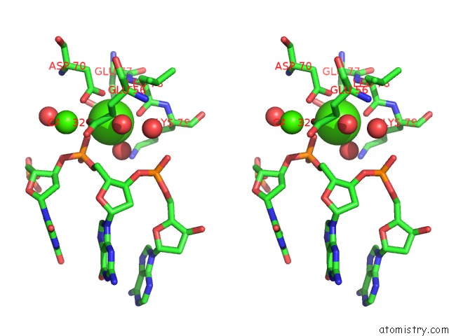
Stereo pair view
Mono view
Stereo pair view
A full contact list of Calcium with other atoms in the Ca binding
site number 2 of Crystal Structure of Muth-Unmethylated Dna Complex within 5.0Å range:
|
Reference:
J.Y.Lee,
J.Chang,
N.Joseph,
R.Ghirlando,
D.N.Rao,
W.Yang.
Muth Complexed with Hemi- and Unmethylated Dnas: Coupling Base Recognition and Dna Cleavage. Mol.Cell V. 20 155 2005.
ISSN: ISSN 1097-2765
PubMed: 16209953
DOI: 10.1016/J.MOLCEL.2005.08.019
Page generated: Tue Jul 8 04:20:33 2025
ISSN: ISSN 1097-2765
PubMed: 16209953
DOI: 10.1016/J.MOLCEL.2005.08.019
Last articles
Mg in 6KI8Mg in 6KJ6
Mg in 6KF9
Mg in 6KE2
Mg in 6KF4
Mg in 6KF3
Mg in 6KE4
Mg in 6KE0
Mg in 6KDZ
Mg in 6KDX