Calcium »
PDB 2agq-2b03 »
2asm »
Calcium in PDB 2asm: Structure of Rabbit Actin in Complex with Reidispongiolide A
Protein crystallography data
The structure of Structure of Rabbit Actin in Complex with Reidispongiolide A, PDB code: 2asm
was solved by
J.S.Allingham,
A.Zampella,
M.V.D'auria,
I.Rayment,
with X-Ray Crystallography technique. A brief refinement statistics is given in the table below:
| Resolution Low / High (Å) | 35.00 / 1.60 |
| Space group | C 1 2 1 |
| Cell size a, b, c (Å), α, β, γ (°) | 171.181, 54.694, 40.674, 90.00, 95.97, 90.00 |
| R / Rfree (%) | 16.4 / 18.9 |
Calcium Binding Sites:
The binding sites of Calcium atom in the Structure of Rabbit Actin in Complex with Reidispongiolide A
(pdb code 2asm). This binding sites where shown within
5.0 Angstroms radius around Calcium atom.
In total 2 binding sites of Calcium where determined in the Structure of Rabbit Actin in Complex with Reidispongiolide A, PDB code: 2asm:
Jump to Calcium binding site number: 1; 2;
In total 2 binding sites of Calcium where determined in the Structure of Rabbit Actin in Complex with Reidispongiolide A, PDB code: 2asm:
Jump to Calcium binding site number: 1; 2;
Calcium binding site 1 out of 2 in 2asm
Go back to
Calcium binding site 1 out
of 2 in the Structure of Rabbit Actin in Complex with Reidispongiolide A
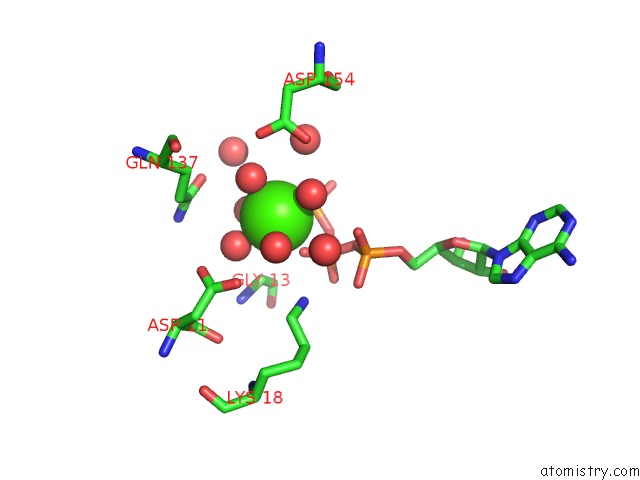
Mono view
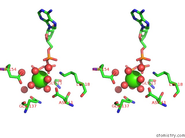
Stereo pair view
Mono view
Stereo pair view
A full contact list of Calcium with other atoms in the Ca binding
site number 1 of Structure of Rabbit Actin in Complex with Reidispongiolide A within 5.0Å range:
|
Calcium binding site 2 out of 2 in 2asm
Go back to
Calcium binding site 2 out
of 2 in the Structure of Rabbit Actin in Complex with Reidispongiolide A
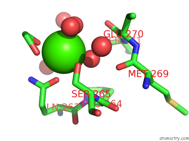
Mono view
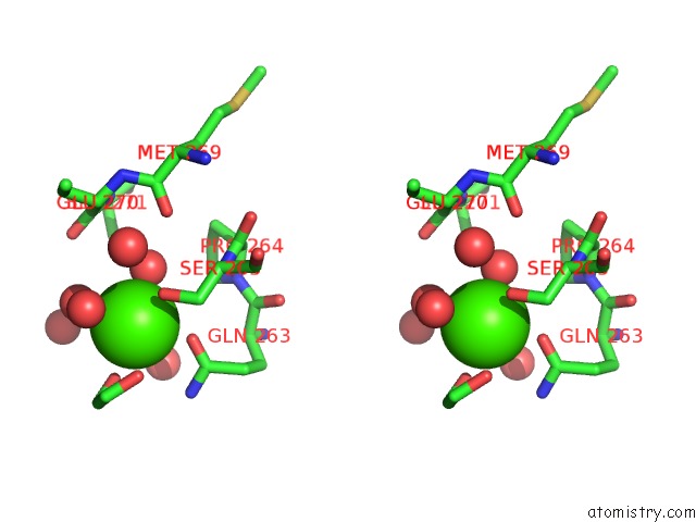
Stereo pair view
Mono view
Stereo pair view
A full contact list of Calcium with other atoms in the Ca binding
site number 2 of Structure of Rabbit Actin in Complex with Reidispongiolide A within 5.0Å range:
|
Reference:
J.S.Allingham,
A.Zampella,
M.V.D'auria,
I.Rayment.
Structures of Microfilament Destabilizing Toxins Bound to Actin Provide Insight Into Toxin Design and Activity Proc.Natl.Acad.Sci.Usa V. 102 14527 2005.
ISSN: ISSN 0027-8424
PubMed: 16192358
DOI: 10.1073/PNAS.0502089102
Page generated: Tue Jul 8 04:22:26 2025
ISSN: ISSN 0027-8424
PubMed: 16192358
DOI: 10.1073/PNAS.0502089102
Last articles
Mg in 6C07Mg in 6BYW
Mg in 6C0J
Mg in 6C06
Mg in 6C05
Mg in 6C04
Mg in 6BZO
Mg in 6BYR
Mg in 6BZ0
Mg in 6BYU