Calcium »
PDB 2c5d-2cn3 »
2c60 »
Calcium in PDB 2c60: Crystal Structure of Human Mitogen-Activated Protein Kinase Kinase Kinase 3 Isoform 2 Phox Domain at 1.25 A Resolution
Enzymatic activity of Crystal Structure of Human Mitogen-Activated Protein Kinase Kinase Kinase 3 Isoform 2 Phox Domain at 1.25 A Resolution
All present enzymatic activity of Crystal Structure of Human Mitogen-Activated Protein Kinase Kinase Kinase 3 Isoform 2 Phox Domain at 1.25 A Resolution:
2.7.1.37;
2.7.1.37;
Protein crystallography data
The structure of Crystal Structure of Human Mitogen-Activated Protein Kinase Kinase Kinase 3 Isoform 2 Phox Domain at 1.25 A Resolution, PDB code: 2c60
was solved by
J.E.Debreczeni,
E.Salah,
E.Papagrigoriou,
N.Burgess,
F.Von Delft,
O.Gileadi,
M.Sundstrom,
A.Edwards,
C.Arrowsmith,
J.Weigelt,
S.Knapp,
with X-Ray Crystallography technique. A brief refinement statistics is given in the table below:
| Resolution Low / High (Å) | 39.72 / 1.25 |
| Space group | C 1 2 1 |
| Cell size a, b, c (Å), α, β, γ (°) | 82.800, 42.300, 30.600, 90.00, 106.60, 90.00 |
| R / Rfree (%) | 16.5 / 18.2 |
Calcium Binding Sites:
The binding sites of Calcium atom in the Crystal Structure of Human Mitogen-Activated Protein Kinase Kinase Kinase 3 Isoform 2 Phox Domain at 1.25 A Resolution
(pdb code 2c60). This binding sites where shown within
5.0 Angstroms radius around Calcium atom.
In total only one binding site of Calcium was determined in the Crystal Structure of Human Mitogen-Activated Protein Kinase Kinase Kinase 3 Isoform 2 Phox Domain at 1.25 A Resolution, PDB code: 2c60:
In total only one binding site of Calcium was determined in the Crystal Structure of Human Mitogen-Activated Protein Kinase Kinase Kinase 3 Isoform 2 Phox Domain at 1.25 A Resolution, PDB code: 2c60:
Calcium binding site 1 out of 1 in 2c60
Go back to
Calcium binding site 1 out
of 1 in the Crystal Structure of Human Mitogen-Activated Protein Kinase Kinase Kinase 3 Isoform 2 Phox Domain at 1.25 A Resolution
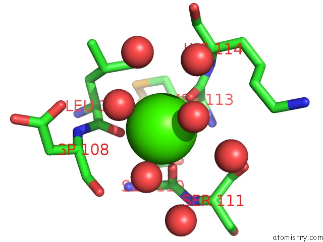
Mono view
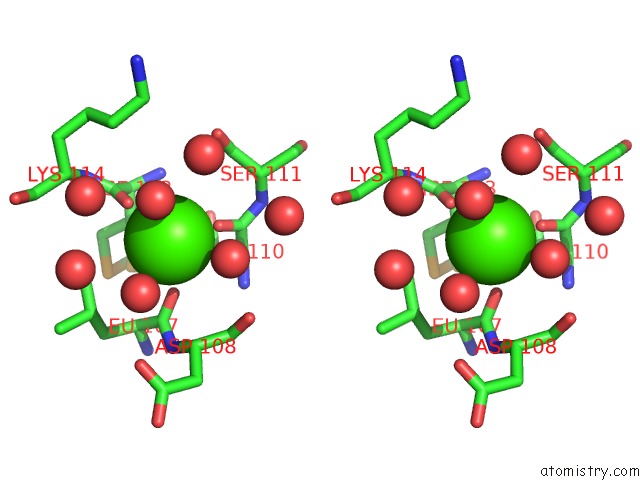
Stereo pair view
Mono view
Stereo pair view
A full contact list of Calcium with other atoms in the Ca binding
site number 1 of Crystal Structure of Human Mitogen-Activated Protein Kinase Kinase Kinase 3 Isoform 2 Phox Domain at 1.25 A Resolution within 5.0Å range:
|
Reference:
J.E.Debreczeni,
E.Salah,
E.Papagrigoriou,
N.Burgess,
F.Von Delft,
O.Gileadi,
M.Sundstrom,
A.Edwards,
C.Arrowsmith,
J.Weigelt,
S.Knapp.
Crystal Structure of Human Mitogen-Activated Protein Kinase Kinase Kinase 3 Isoform 2 Fox Domain at 1.25 A Resolution To Be Published.
Page generated: Tue Jul 8 04:46:53 2025
Last articles
Mg in 4Z1IMg in 4Z17
Mg in 4YPO
Mg in 4YZM
Mg in 4Z0V
Mg in 4YZD
Mg in 4YXW
Mg in 4YWE
Mg in 4YWS
Mg in 4YSJ