Calcium »
PDB 2cn6-2dbx »
2d3d »
Calcium in PDB 2d3d: Crystal Structure of the Rna Binding Sam Domain of Saccharomyces Cerevisiae VTS1
Protein crystallography data
The structure of Crystal Structure of the Rna Binding Sam Domain of Saccharomyces Cerevisiae VTS1, PDB code: 2d3d
was solved by
T.Aviv,
A.N.Amborski,
X.S.Zhao,
J.J.Kwan,
P.E.Johnson,
F.Sicheri,
L.W.Donaldson,
with X-Ray Crystallography technique. A brief refinement statistics is given in the table below:
| Resolution Low / High (Å) | 26.60 / 1.60 |
| Space group | P 21 21 21 |
| Cell size a, b, c (Å), α, β, γ (°) | 27.380, 27.890, 99.910, 90.00, 90.00, 90.00 |
| R / Rfree (%) | 20.6 / 25.6 |
Calcium Binding Sites:
The binding sites of Calcium atom in the Crystal Structure of the Rna Binding Sam Domain of Saccharomyces Cerevisiae VTS1
(pdb code 2d3d). This binding sites where shown within
5.0 Angstroms radius around Calcium atom.
In total only one binding site of Calcium was determined in the Crystal Structure of the Rna Binding Sam Domain of Saccharomyces Cerevisiae VTS1, PDB code: 2d3d:
In total only one binding site of Calcium was determined in the Crystal Structure of the Rna Binding Sam Domain of Saccharomyces Cerevisiae VTS1, PDB code: 2d3d:
Calcium binding site 1 out of 1 in 2d3d
Go back to
Calcium binding site 1 out
of 1 in the Crystal Structure of the Rna Binding Sam Domain of Saccharomyces Cerevisiae VTS1
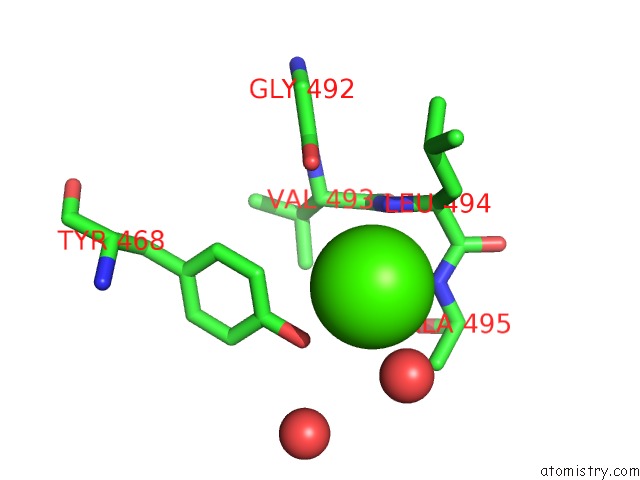
Mono view
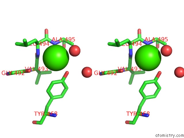
Stereo pair view
Mono view
Stereo pair view
A full contact list of Calcium with other atoms in the Ca binding
site number 1 of Crystal Structure of the Rna Binding Sam Domain of Saccharomyces Cerevisiae VTS1 within 5.0Å range:
|
Reference:
T.Aviv,
A.N.Amborski,
X.S.Zhao,
J.J.Kwan,
P.E.Johnson,
F.Sicheri,
L.W.Donaldson.
The uc(Nmr) and X-Ray Structures of the Saccharomyces Cerevisiae VTS1 Sam Domain Define A Surface For the Recognition of Rna Hairpins J.Mol.Biol. V. 356 274 2006.
ISSN: ISSN 0022-2836
PubMed: 16375924
DOI: 10.1016/J.JMB.2005.11.066
Page generated: Tue Jul 8 04:58:13 2025
ISSN: ISSN 0022-2836
PubMed: 16375924
DOI: 10.1016/J.JMB.2005.11.066
Last articles
Ir in 3PK2Ir in 3F2T
Ir in 3IRW
Ir in 3D0U
Ir in 2GIS
Ir in 3B31
Ir in 1K26
Ir in 2B0U
Ir in 1C1K
Ir in 1ICG