Calcium »
PDB 2cn6-2dbx »
2d7f »
Calcium in PDB 2d7f: Crystal Structure of A Lectin From Canavalia Gladiata Seeds Complexed with Alpha-Methyl-Mannoside and Alpha- Aminobutyric Acid
Protein crystallography data
The structure of Crystal Structure of A Lectin From Canavalia Gladiata Seeds Complexed with Alpha-Methyl-Mannoside and Alpha- Aminobutyric Acid, PDB code: 2d7f
was solved by
P.Delatorre,
B.A.M.Rocha,
E.P.Souza,
B.T.Freitas,
F.B.B.M.Moreno,
A.H.Sampaio,
W.F.Azevedo Jr.,
B.S.Cavada,
with X-Ray Crystallography technique. A brief refinement statistics is given in the table below:
| Resolution Low / High (Å) | 9.99 / 2.31 |
| Space group | C 2 2 21 |
| Cell size a, b, c (Å), α, β, γ (°) | 100.915, 115.754, 241.626, 90.00, 90.00, 90.00 |
| R / Rfree (%) | 18 / 22.7 |
Other elements in 2d7f:
The structure of Crystal Structure of A Lectin From Canavalia Gladiata Seeds Complexed with Alpha-Methyl-Mannoside and Alpha- Aminobutyric Acid also contains other interesting chemical elements:
| Manganese | (Mn) | 4 atoms |
Calcium Binding Sites:
The binding sites of Calcium atom in the Crystal Structure of A Lectin From Canavalia Gladiata Seeds Complexed with Alpha-Methyl-Mannoside and Alpha- Aminobutyric Acid
(pdb code 2d7f). This binding sites where shown within
5.0 Angstroms radius around Calcium atom.
In total 4 binding sites of Calcium where determined in the Crystal Structure of A Lectin From Canavalia Gladiata Seeds Complexed with Alpha-Methyl-Mannoside and Alpha- Aminobutyric Acid, PDB code: 2d7f:
Jump to Calcium binding site number: 1; 2; 3; 4;
In total 4 binding sites of Calcium where determined in the Crystal Structure of A Lectin From Canavalia Gladiata Seeds Complexed with Alpha-Methyl-Mannoside and Alpha- Aminobutyric Acid, PDB code: 2d7f:
Jump to Calcium binding site number: 1; 2; 3; 4;
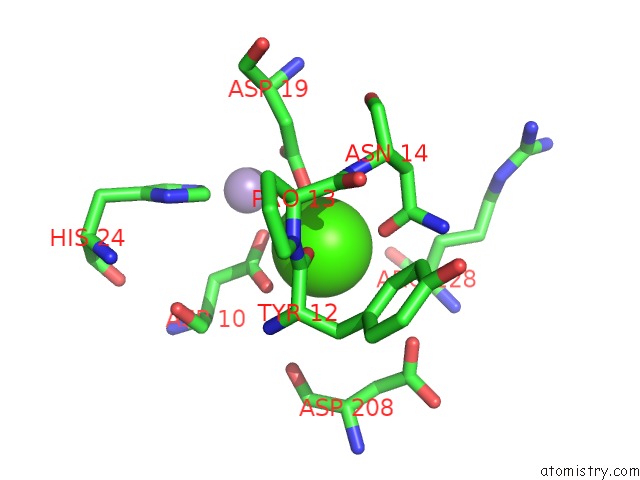
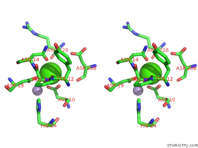
Calcium binding site 1 out of 4 in 2d7f
Go back to
Calcium binding site 1 out
of 4 in the Crystal Structure of A Lectin From Canavalia Gladiata Seeds Complexed with Alpha-Methyl-Mannoside and Alpha- Aminobutyric Acid
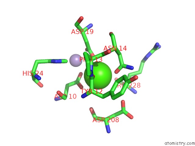
Mono view
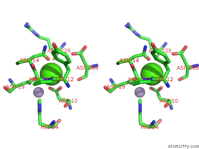
Stereo pair view
Mono view
Stereo pair view
A full contact list of Calcium with other atoms in the Ca binding
site number 1 of Crystal Structure of A Lectin From Canavalia Gladiata Seeds Complexed with Alpha-Methyl-Mannoside and Alpha- Aminobutyric Acid within 5.0Å range:
|
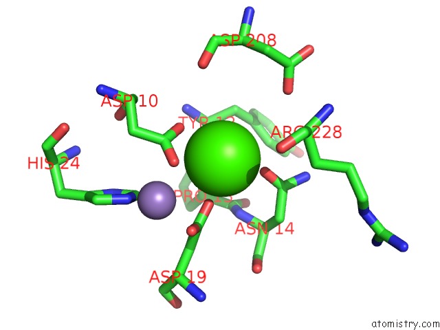
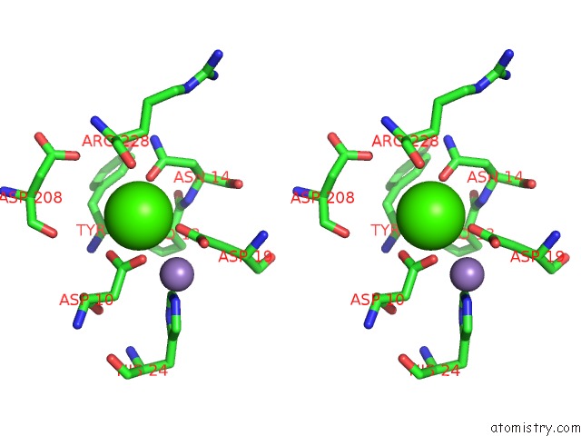
Calcium binding site 2 out of 4 in 2d7f
Go back to
Calcium binding site 2 out
of 4 in the Crystal Structure of A Lectin From Canavalia Gladiata Seeds Complexed with Alpha-Methyl-Mannoside and Alpha- Aminobutyric Acid
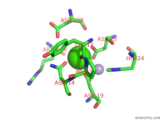
Mono view
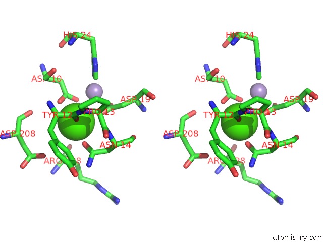
Stereo pair view
Mono view
Stereo pair view
A full contact list of Calcium with other atoms in the Ca binding
site number 2 of Crystal Structure of A Lectin From Canavalia Gladiata Seeds Complexed with Alpha-Methyl-Mannoside and Alpha- Aminobutyric Acid within 5.0Å range:
|
Calcium binding site 3 out of 4 in 2d7f
Go back to
Calcium binding site 3 out
of 4 in the Crystal Structure of A Lectin From Canavalia Gladiata Seeds Complexed with Alpha-Methyl-Mannoside and Alpha- Aminobutyric Acid
Mono view
Stereo pair view
Mono view
Stereo pair view
A full contact list of Calcium with other atoms in the Ca binding
site number 3 of Crystal Structure of A Lectin From Canavalia Gladiata Seeds Complexed with Alpha-Methyl-Mannoside and Alpha- Aminobutyric Acid within 5.0Å range:
|
Calcium binding site 4 out of 4 in 2d7f
Go back to
Calcium binding site 4 out
of 4 in the Crystal Structure of A Lectin From Canavalia Gladiata Seeds Complexed with Alpha-Methyl-Mannoside and Alpha- Aminobutyric Acid
Mono view
Stereo pair view
Mono view
Stereo pair view
A full contact list of Calcium with other atoms in the Ca binding
site number 4 of Crystal Structure of A Lectin From Canavalia Gladiata Seeds Complexed with Alpha-Methyl-Mannoside and Alpha- Aminobutyric Acid within 5.0Å range:
|
Reference:
P.Delatorre,
B.A.M.Rocha,
E.P.Souza,
T.M.Oliveira,
G.A.Bezerra,
F.B.M.B.Moreno,
B.T.Freitas,
T.Santi-Gadelha,
A.H.Sampaio,
W.F.Azevedo Jr.,
B.S.Cavada.
Structure of A Lectin From Canavalia Gladiata Seeds: New Structural Insights For Old Molecules Bmc Struct.Biol. V. 7 52 2007.
ISSN: ESSN 1472-6807
PubMed: 17683532
DOI: 10.1186/1472-6807-7-52
Page generated: Tue Jul 8 05:00:02 2025
ISSN: ESSN 1472-6807
PubMed: 17683532
DOI: 10.1186/1472-6807-7-52
Last articles
I in 6SPWI in 6S42
I in 6SQ4
I in 6S4L
I in 6S0T
I in 6S1X
I in 6P5T
I in 6PKG
I in 6RNS
I in 6RZM