Atomistry » Calcium » PDB 2dcj-2dw0 » 2ddr
Atomistry »
  Calcium »
    PDB 2dcj-2dw0 »
      2ddr »

Calcium in PDB 2ddr: Crystal Structure of Sphingomyelinase From Bacillus Cereus with Calcium Ion

Enzymatic activity of Crystal Structure of Sphingomyelinase From Bacillus Cereus with Calcium Ion

All present enzymatic activity of Crystal Structure of Sphingomyelinase From Bacillus Cereus with Calcium Ion:
3.1.4.12;

Protein crystallography data

The structure of Crystal Structure of Sphingomyelinase From Bacillus Cereus with Calcium Ion, PDB code: 2ddr was solved by H.Ago, M.Oda, M.Takahashi, H.Tsuge, S.Ochi, N.Katunuma, M.Miyano, J.Sakurai, Riken Structural Genomics/Proteomics Initiative(Rsgi), with X-Ray Crystallography technique. A brief refinement statistics is given in the table below:

Resolution Low / High (Å) 19.71 / 1.40
Space group P 1
Cell size a, b, c (Å), α, β, γ (°) 65.430, 72.691, 77.859, 112.19, 89.97, 116.76
R / Rfree (%) 21.3 / 23.4

Calcium Binding Sites:

The binding sites of Calcium atom in the Crystal Structure of Sphingomyelinase From Bacillus Cereus with Calcium Ion (pdb code 2ddr). This binding sites where shown within 5.0 Angstroms radius around Calcium atom.
In total 8 binding sites of Calcium where determined in the Crystal Structure of Sphingomyelinase From Bacillus Cereus with Calcium Ion, PDB code: 2ddr:
Jump to Calcium binding site number: 1; 2; 3; 4; 5; 6; 7; 8;

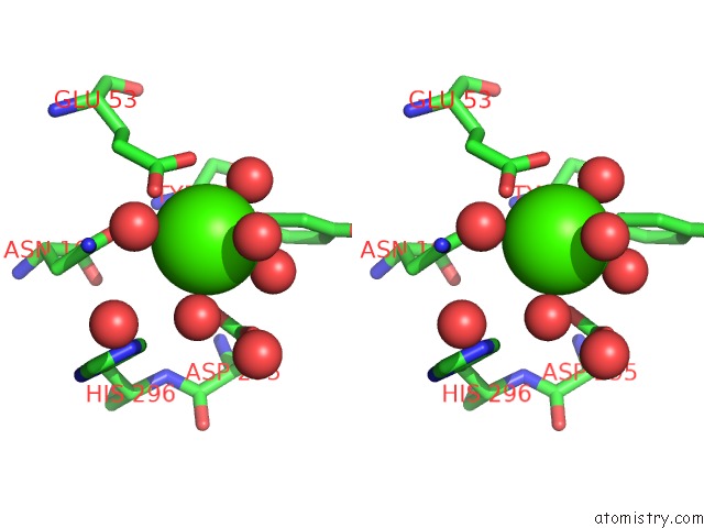
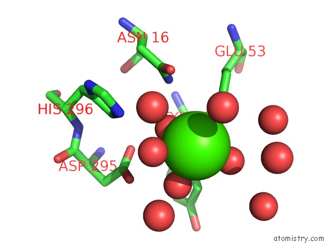
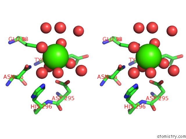
Calcium binding site 1 out of 8 in 2ddr

Go back to Calcium Binding Sites List in 2ddr
Calcium binding site 1 out of 8 in the Crystal Structure of Sphingomyelinase From Bacillus Cereus with Calcium Ion


Mono view


Stereo pair view

A full contact list of Calcium with other atoms in the Ca binding site number 1 of Crystal Structure of Sphingomyelinase From Bacillus Cereus with Calcium Ion within 5.0Å range:
probe atom residue distance (Å) B Occ
A:Ca1324

b:10.4
occ:1.00
OD1 A:ASN57 2.3 11.2 1.0
O A:PHE55 2.3 10.8 1.0
OE1 A:GLU99 2.3 11.8 1.0
OD2 A:ASP100 2.3 14.2 1.0
O A:HOH1331 2.3 12.1 1.0
O A:HOH1345 2.4 15.1 1.0
CG A:ASP100 3.3 12.5 1.0
CD A:GLU99 3.3 10.6 1.0
C A:PHE55 3.4 8.9 1.0
CG A:ASN57 3.4 11.4 1.0
OE2 A:GLU99 3.7 13.7 1.0
NH2 A:ARG80 3.8 13.2 1.0
OD1 A:ASP100 3.9 13.6 1.0
NE A:ARG80 4.0 11.6 1.0
N A:ASN57 4.1 10.5 1.0
CA A:PHE55 4.1 9.1 1.0
ND2 A:ASN57 4.1 15.0 1.0
N A:ASP100 4.2 8.7 1.0
O A:HOH1394 4.2 21.4 1.0
CZ A:ARG80 4.2 13.0 1.0
CB A:ASP100 4.3 10.8 1.0
O A:HOH1400 4.4 21.6 1.0
N A:ASP56 4.5 9.7 1.0
C A:ASP56 4.5 10.1 1.0
CB A:ASN57 4.6 10.5 1.0
CA A:ASN57 4.6 9.6 1.0
CG A:GLU99 4.7 9.8 1.0
CA A:ASP56 4.7 10.1 1.0
CB A:PHE55 4.7 9.8 1.0
CA A:ASP100 4.9 10.0 1.0
O A:HOH1376 4.9 19.8 1.0
CA A:GLU99 5.0 10.8 1.0
CD A:ARG80 5.0 13.0 1.0

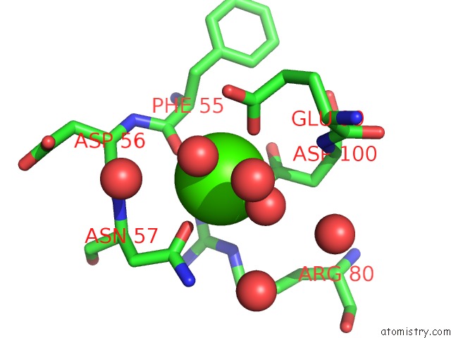
Calcium binding site 2 out of 8 in 2ddr

Go back to Calcium Binding Sites List in 2ddr
Calcium binding site 2 out of 8 in the Crystal Structure of Sphingomyelinase From Bacillus Cereus with Calcium Ion


Mono view


Stereo pair view

A full contact list of Calcium with other atoms in the Ca binding site number 2 of Crystal Structure of Sphingomyelinase From Bacillus Cereus with Calcium Ion within 5.0Å range:
probe atom residue distance (Å) B Occ
A:Ca1325

b:20.8
occ:1.00
OE1 A:GLU53 2.4 13.6 1.0
O A:HOH1387 2.4 20.4 1.0
O A:HOH1333 2.4 13.6 1.0
O A:HOH1380 2.5 20.0 1.0
O A:HOH1479 2.5 28.3 1.0
O A:HOH1492 2.8 30.0 1.0
CD A:GLU53 3.2 12.9 1.0
OE2 A:GLU53 3.4 15.1 1.0
O A:HOH1447 3.8 25.4 1.0
ND2 A:ASN16 4.2 10.5 1.0
OD2 A:ASP295 4.2 15.4 1.0
CD1 A:TYR18 4.4 11.2 1.0
NE2 A:HIS296 4.4 13.9 1.0
O A:HOH1372 4.5 19.1 1.0
OD1 A:ASP295 4.6 13.3 1.0
CD2 A:HIS296 4.6 14.3 1.0
CG A:GLU53 4.6 10.8 1.0
OD1 A:ASN16 4.6 9.1 1.0
CE1 A:TYR18 4.7 12.6 1.0
O A:HOH1623 4.7 43.4 1.0
CG A:ASN16 4.8 8.1 1.0
CG A:ASP295 4.9 14.4 1.0

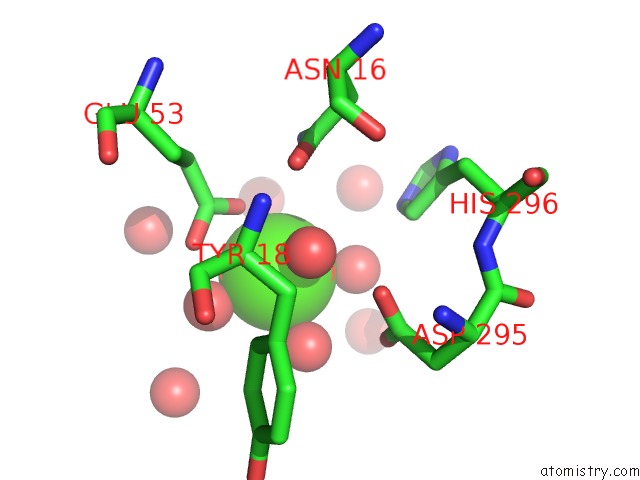
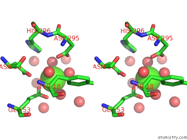
Calcium binding site 3 out of 8 in 2ddr

Go back to Calcium Binding Sites List in 2ddr
Calcium binding site 3 out of 8 in the Crystal Structure of Sphingomyelinase From Bacillus Cereus with Calcium Ion


Mono view


Stereo pair view

A full contact list of Calcium with other atoms in the Ca binding site number 3 of Crystal Structure of Sphingomyelinase From Bacillus Cereus with Calcium Ion within 5.0Å range:
probe atom residue distance (Å) B Occ
B:Ca1326

b:10.9
occ:1.00
O B:PHE55 2.3 10.2 1.0
OD2 B:ASP100 2.3 14.4 1.0
OD1 B:ASN57 2.3 11.7 1.0
OE1 B:GLU99 2.3 11.6 1.0
O B:HOH1335 2.4 13.1 1.0
O B:HOH1357 2.4 16.8 1.0
CG B:ASP100 3.3 11.8 1.0
CD B:GLU99 3.3 10.8 1.0
C B:PHE55 3.4 9.5 1.0
CG B:ASN57 3.5 13.4 1.0
OE2 B:GLU99 3.7 13.0 1.0
NH2 B:ARG80 3.9 13.0 1.0
OD1 B:ASP100 3.9 11.3 1.0
NE B:ARG80 4.0 12.7 1.0
N B:ASN57 4.1 11.9 1.0
CA B:PHE55 4.1 8.8 1.0
N B:ASP100 4.2 9.2 1.0
ND2 B:ASN57 4.2 15.0 1.0
CB B:ASP100 4.3 9.8 1.0
O B:HOH1485 4.3 26.2 1.0
CZ B:ARG80 4.3 13.6 1.0
O B:HOH1407 4.3 21.4 1.0
N B:ASP56 4.5 9.2 1.0
C B:ASP56 4.5 10.7 1.0
CB B:ASN57 4.6 12.1 1.0
CA B:ASN57 4.6 11.2 1.0
CA B:ASP56 4.6 9.1 1.0
CG B:GLU99 4.7 11.8 1.0
CB B:PHE55 4.7 9.4 1.0
CA B:ASP100 4.8 9.4 1.0
O B:HOH1415 4.9 21.5 1.0
CA B:GLU99 5.0 11.5 1.0

Calcium binding site 4 out of 8 in 2ddr

Go back to Calcium Binding Sites List in 2ddr
Calcium binding site 4 out of 8 in the Crystal Structure of Sphingomyelinase From Bacillus Cereus with Calcium Ion


Mono view


Stereo pair view

A full contact list of Calcium with other atoms in the Ca binding site number 4 of Crystal Structure of Sphingomyelinase From Bacillus Cereus with Calcium Ion within 5.0Å range:
probe atom residue distance (Å) B Occ
B:Ca1327

b:19.6
occ:1.00
OE1 B:GLU53 2.3 14.2 1.0
O B:HOH1441 2.4 23.0 1.0
O B:HOH1403 2.5 20.8 1.0
O B:HOH1343 2.5 13.7 1.0
O B:HOH1482 2.6 26.8 1.0
O B:HOH1493 2.7 28.2 1.0
CD B:GLU53 3.2 12.4 1.0
OE2 B:GLU53 3.4 15.7 1.0
O B:HOH1453 3.8 25.2 1.0
O B:HOH1438 4.2 23.8 1.0
ND2 B:ASN16 4.2 11.4 1.0
OD2 B:ASP295 4.3 16.4 1.0
CD1 B:TYR18 4.4 11.8 1.0
NE2 B:HIS296 4.4 12.1 1.0
O B:HOH1351 4.5 15.5 1.0
CG B:GLU53 4.6 10.7 1.0
OD1 B:ASN16 4.7 10.6 1.0
CD2 B:HIS296 4.7 13.2 1.0
CE1 B:TYR18 4.7 13.0 1.0
OD1 B:ASP295 4.7 14.1 1.0
CG B:ASN16 4.9 10.2 1.0
CG B:ASP295 5.0 13.1 1.0

Calcium binding site 5 out of 8 in 2ddr

Go back to Calcium Binding Sites List in 2ddr
Calcium binding site 5 out of 8 in the Crystal Structure of Sphingomyelinase From Bacillus Cereus with Calcium Ion


Mono view


Stereo pair view

A full contact list of Calcium with other atoms in the Ca binding site number 5 of Crystal Structure of Sphingomyelinase From Bacillus Cereus with Calcium Ion within 5.0Å range:
probe atom residue distance (Å) B Occ
C:Ca1328

b:10.2
occ:1.00
O C:PHE55 2.3 10.3 1.0
OD2 C:ASP100 2.3 14.5 1.0
OD1 C:ASN57 2.3 12.2 1.0
OE1 C:GLU99 2.3 13.4 1.0
O C:HOH1337 2.3 13.3 1.0
O C:HOH1332 2.4 11.1 1.0
CG C:ASP100 3.2 13.4 1.0
CD C:GLU99 3.3 11.6 1.0
C C:PHE55 3.4 8.1 1.0
CG C:ASN57 3.5 12.3 1.0
OE2 C:GLU99 3.7 13.2 1.0
NH2 C:ARG80 3.8 12.7 1.0
OD1 C:ASP100 3.9 13.6 1.0
NE C:ARG80 4.0 12.5 1.0
N C:ASN57 4.1 9.3 1.0
CA C:PHE55 4.1 9.2 1.0
ND2 C:ASN57 4.1 15.6 1.0
N C:ASP100 4.2 8.4 1.0
O C:HOH1376 4.2 18.9 1.0
CZ C:ARG80 4.2 12.7 1.0
CB C:ASP100 4.2 10.7 1.0
O C:HOH1355 4.3 15.9 1.0
N C:ASP56 4.5 8.7 1.0
C C:ASP56 4.5 9.6 1.0
CB C:ASN57 4.6 10.2 1.0
O C:HOH1570 4.6 41.0 1.0
CA C:ASN57 4.6 9.7 1.0
CA C:ASP56 4.7 9.2 1.0
CB C:PHE55 4.7 8.9 1.0
CG C:GLU99 4.7 9.7 1.0
O C:HOH1363 4.8 16.6 1.0
CA C:ASP100 4.8 9.9 1.0
CD C:ARG80 5.0 13.3 1.0

Calcium binding site 6 out of 8 in 2ddr

Go back to Calcium Binding Sites List in 2ddr
Calcium binding site 6 out of 8 in the Crystal Structure of Sphingomyelinase From Bacillus Cereus with Calcium Ion


Mono view


Stereo pair view

A full contact list of Calcium with other atoms in the Ca binding site number 6 of Crystal Structure of Sphingomyelinase From Bacillus Cereus with Calcium Ion within 5.0Å range:
probe atom residue distance (Å) B Occ
C:Ca1329

b:20.9
occ:1.00
O C:HOH1390 2.3 21.0 1.0
OE2 C:GLU53 2.4 14.5 1.0
O C:HOH1341 2.4 14.0 1.0
O C:HOH1397 2.4 21.0 1.0
O C:HOH1464 2.5 30.9 1.0
CD C:GLU53 3.3 13.7 1.0
OE1 C:GLU53 3.4 14.8 1.0
O C:HOH1411 4.0 22.6 1.0
OD2 C:ASP295 4.2 15.4 1.0
ND2 C:ASN16 4.2 11.0 1.0
NE2 C:HIS296 4.3 14.4 1.0
CD1 C:TYR18 4.4 11.4 1.0
O C:HOH1567 4.4 31.4 1.0
CD2 C:HIS296 4.6 14.3 1.0
O C:HOH1364 4.6 17.0 1.0
OD1 C:ASP295 4.6 13.2 1.0
OD1 C:ASN16 4.6 9.5 1.0
CE1 C:TYR18 4.7 12.5 1.0
CG C:GLU53 4.7 10.2 1.0
O C:HOH1513 4.8 47.2 1.0
O C:HOH1439 4.8 25.8 1.0
CG C:ASN16 4.8 8.8 1.0
CG C:ASP295 4.8 14.7 1.0

Calcium binding site 7 out of 8 in 2ddr

Go back to Calcium Binding Sites List in 2ddr
Calcium binding site 7 out of 8 in the Crystal Structure of Sphingomyelinase From Bacillus Cereus with Calcium Ion


Mono view


Stereo pair view

A full contact list of Calcium with other atoms in the Ca binding site number 7 of Crystal Structure of Sphingomyelinase From Bacillus Cereus with Calcium Ion within 5.0Å range:
probe atom residue distance (Å) B Occ
D:Ca1330

b:11.2
occ:1.00
OD2 D:ASP100 2.2 13.9 1.0
OD1 D:ASN57 2.3 11.9 1.0
OE1 D:GLU99 2.3 13.6 1.0
O D:PHE55 2.3 10.7 1.0
O D:HOH1348 2.4 15.1 1.0
O D:HOH1338 2.4 12.8 1.0
CG D:ASP100 3.3 12.0 1.0
CD D:GLU99 3.3 12.0 1.0
C D:PHE55 3.4 10.2 1.0
CG D:ASN57 3.4 13.6 1.0
OE2 D:GLU99 3.7 12.8 1.0
NH2 D:ARG80 3.9 12.4 1.0
OD1 D:ASP100 3.9 12.2 1.0
NE D:ARG80 4.0 10.7 1.0
N D:ASN57 4.1 11.7 1.0
CA D:PHE55 4.1 9.3 1.0
N D:ASP100 4.1 8.9 1.0
ND2 D:ASN57 4.2 14.9 1.0
CB D:ASP100 4.2 10.0 1.0
O D:HOH1429 4.2 24.8 1.0
CZ D:ARG80 4.3 12.5 1.0
O D:HOH1384 4.4 20.1 1.0
N D:ASP56 4.5 9.5 1.0
C D:ASP56 4.5 10.6 1.0
CB D:ASN57 4.5 11.7 1.0
CA D:ASN57 4.5 11.1 1.0
CG D:GLU99 4.6 10.8 1.0
CA D:ASP56 4.7 9.3 1.0
CB D:PHE55 4.7 9.7 1.0
CA D:ASP100 4.8 9.9 1.0
O D:HOH1389 4.9 19.1 1.0
CA D:GLU99 5.0 10.3 1.0

Calcium binding site 8 out of 8 in 2ddr

Go back to Calcium Binding Sites List in 2ddr
Calcium binding site 8 out of 8 in the Crystal Structure of Sphingomyelinase From Bacillus Cereus with Calcium Ion


Mono view


Stereo pair view

A full contact list of Calcium with other atoms in the Ca binding site number 8 of Crystal Structure of Sphingomyelinase From Bacillus Cereus with Calcium Ion within 5.0Å range:
probe atom residue distance (Å) B Occ
D:Ca1331

b:20.1
occ:1.00
O D:HOH1397 2.4 20.6 1.0
OE1 D:GLU53 2.4 14.7 1.0
O D:HOH1346 2.5 14.0 1.0
O D:HOH1460 2.5 29.5 1.0
O D:HOH1427 2.5 24.3 1.0
CD D:GLU53 3.3 13.4 1.0
OE2 D:GLU53 3.4 15.3 1.0
O D:HOH1455 3.9 26.6 1.0
O D:HOH1633 4.2 29.6 1.0
OD1 D:ASN16 4.2 13.9 1.0
OD2 D:ASP295 4.3 16.5 1.0
NE2 D:HIS296 4.3 11.4 1.0
CD1 D:TYR18 4.4 11.3 1.0
O D:HOH1360 4.5 16.4 1.0
O D:HOH1509 4.6 52.1 1.0
CD2 D:HIS296 4.7 13.0 1.0
CG D:GLU53 4.7 10.6 1.0
CE1 D:TYR18 4.7 12.9 1.0
OD1 D:ASP295 4.7 13.5 1.0
ND2 D:ASN16 4.7 7.5 1.0
CG D:ASN16 4.9 10.8 1.0
O D:HOH1517 4.9 52.9 1.0
CG D:ASP295 4.9 13.8 1.0
O D:HOH1470 5.0 31.2 1.0

Reference:

H.Ago, M.Oda, M.Takahashi, H.Tsuge, S.Ochi, N.Katunuma, M.Miyano, J.Sakurai. Structural Basis of the Sphingomyelin Phosphodiesterase Activity in Neutral Sphingomyelinase From Bacillus Cereus. J.Biol.Chem. V. 281 16157 2006.
ISSN: ISSN 0021-9258
PubMed: 16595670
DOI: 10.1074/JBC.M601089200
Page generated: Tue Jul 8 05:00:42 2025

Last articles

Mg in 4JI1
Mg in 4JI0
Mg in 4JI2
Mg in 4JI3
Mg in 4JHD
Mg in 4JH6
Mg in 4JH8
Mg in 4JH7
Mg in 4JH3
Mg in 4JH5
© Copyright 2008-2020 by atomistry.com
Home   |    Site Map   |    Copyright   |    Contact us   |    Privacy