Calcium »
PDB 2dcj-2dw0 »
2dob »
Calcium in PDB 2dob: Crystal Structure of Human Saposin A
Protein crystallography data
The structure of Crystal Structure of Human Saposin A, PDB code: 2dob
was solved by
G.G.Prive,
V.E.Ahn,
with X-Ray Crystallography technique. A brief refinement statistics is given in the table below:
| Resolution Low / High (Å) | 27.18 / 2.00 |
| Space group | P 21 21 2 |
| Cell size a, b, c (Å), α, β, γ (°) | 45.680, 50.040, 33.820, 90.00, 90.00, 90.00 |
| R / Rfree (%) | 21.8 / 26.7 |
Calcium Binding Sites:
The binding sites of Calcium atom in the Crystal Structure of Human Saposin A
(pdb code 2dob). This binding sites where shown within
5.0 Angstroms radius around Calcium atom.
In total only one binding site of Calcium was determined in the Crystal Structure of Human Saposin A, PDB code: 2dob:
In total only one binding site of Calcium was determined in the Crystal Structure of Human Saposin A, PDB code: 2dob:
Calcium binding site 1 out of 1 in 2dob
Go back to
Calcium binding site 1 out
of 1 in the Crystal Structure of Human Saposin A
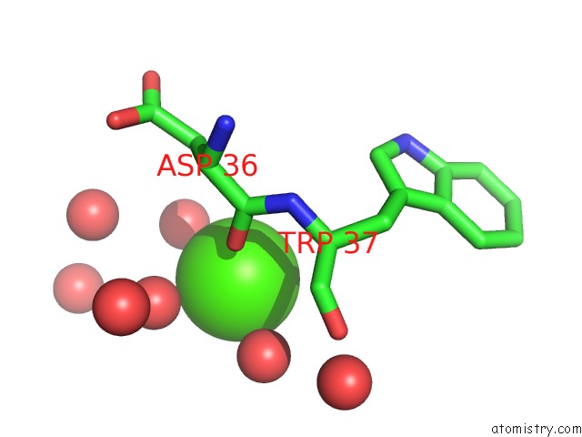
Mono view
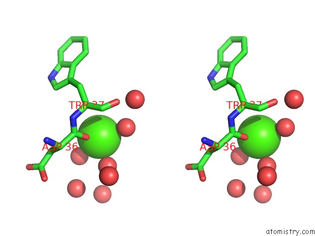
Stereo pair view
Mono view
Stereo pair view
A full contact list of Calcium with other atoms in the Ca binding
site number 1 of Crystal Structure of Human Saposin A within 5.0Å range:
|
Reference:
V.E.Ahn,
P.Leyko,
J.R.Alattia,
L.Chen,
G.G.Prive.
Crystal Structures of Saposins A and C. Protein Sci. V. 15 1849 2006.
ISSN: ISSN 0961-8368
PubMed: 16823039
DOI: 10.1110/PS.062256606
Page generated: Tue Jul 8 05:04:19 2025
ISSN: ISSN 0961-8368
PubMed: 16823039
DOI: 10.1110/PS.062256606
Last articles
Mg in 4JI1Mg in 4JI0
Mg in 4JI2
Mg in 4JI3
Mg in 4JHD
Mg in 4JH6
Mg in 4JH8
Mg in 4JH7
Mg in 4JH3
Mg in 4JH5