Calcium »
PDB 2dcj-2dw0 »
2dvf »
Calcium in PDB 2dvf: Crystals of Peanut Lectin Grown in the Presence of Gal- Alpha-1,3-Gal-Beta-1,4-Gal
Protein crystallography data
The structure of Crystals of Peanut Lectin Grown in the Presence of Gal- Alpha-1,3-Gal-Beta-1,4-Gal, PDB code: 2dvf
was solved by
S.K.Natchiar,
O.Srinivas,
N.Mitra,
A.Surolia,
N.Jayaraman,
M.Vijayan,
with X-Ray Crystallography technique. A brief refinement statistics is given in the table below:
| Resolution Low / High (Å) | 19.94 / 2.74 |
| Space group | P 21 21 2 |
| Cell size a, b, c (Å), α, β, γ (°) | 125.810, 124.060, 75.400, 90.00, 90.00, 90.00 |
| R / Rfree (%) | 20 / 26.3 |
Other elements in 2dvf:
The structure of Crystals of Peanut Lectin Grown in the Presence of Gal- Alpha-1,3-Gal-Beta-1,4-Gal also contains other interesting chemical elements:
| Manganese | (Mn) | 4 atoms |
Calcium Binding Sites:
The binding sites of Calcium atom in the Crystals of Peanut Lectin Grown in the Presence of Gal- Alpha-1,3-Gal-Beta-1,4-Gal
(pdb code 2dvf). This binding sites where shown within
5.0 Angstroms radius around Calcium atom.
In total 4 binding sites of Calcium where determined in the Crystals of Peanut Lectin Grown in the Presence of Gal- Alpha-1,3-Gal-Beta-1,4-Gal, PDB code: 2dvf:
Jump to Calcium binding site number: 1; 2; 3; 4;
In total 4 binding sites of Calcium where determined in the Crystals of Peanut Lectin Grown in the Presence of Gal- Alpha-1,3-Gal-Beta-1,4-Gal, PDB code: 2dvf:
Jump to Calcium binding site number: 1; 2; 3; 4;
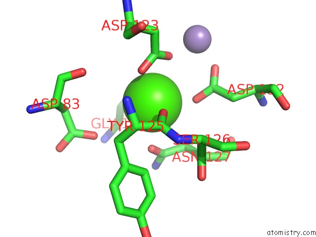
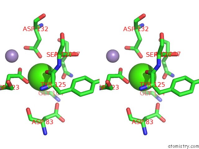
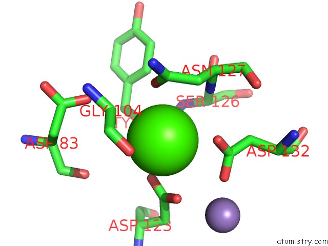
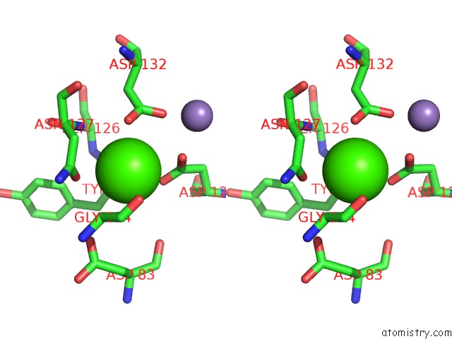
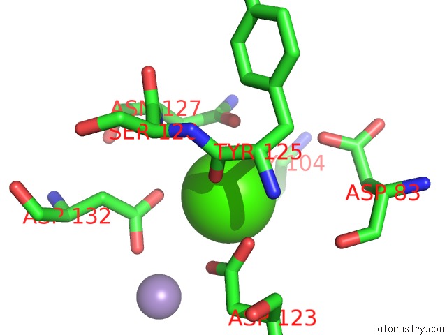
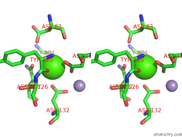
Calcium binding site 1 out of 4 in 2dvf
Go back to
Calcium binding site 1 out
of 4 in the Crystals of Peanut Lectin Grown in the Presence of Gal- Alpha-1,3-Gal-Beta-1,4-Gal
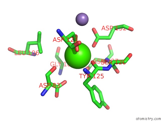
Mono view
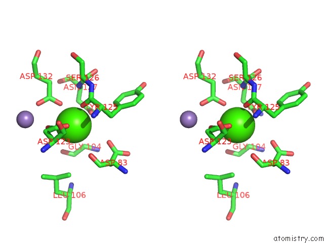
Stereo pair view
Mono view
Stereo pair view
A full contact list of Calcium with other atoms in the Ca binding
site number 1 of Crystals of Peanut Lectin Grown in the Presence of Gal- Alpha-1,3-Gal-Beta-1,4-Gal within 5.0Å range:
|
Calcium binding site 2 out of 4 in 2dvf
Go back to
Calcium binding site 2 out
of 4 in the Crystals of Peanut Lectin Grown in the Presence of Gal- Alpha-1,3-Gal-Beta-1,4-Gal
Mono view
Stereo pair view
Mono view
Stereo pair view
A full contact list of Calcium with other atoms in the Ca binding
site number 2 of Crystals of Peanut Lectin Grown in the Presence of Gal- Alpha-1,3-Gal-Beta-1,4-Gal within 5.0Å range:
|
Calcium binding site 3 out of 4 in 2dvf
Go back to
Calcium binding site 3 out
of 4 in the Crystals of Peanut Lectin Grown in the Presence of Gal- Alpha-1,3-Gal-Beta-1,4-Gal
Mono view
Stereo pair view
Mono view
Stereo pair view
A full contact list of Calcium with other atoms in the Ca binding
site number 3 of Crystals of Peanut Lectin Grown in the Presence of Gal- Alpha-1,3-Gal-Beta-1,4-Gal within 5.0Å range:
|
Calcium binding site 4 out of 4 in 2dvf
Go back to
Calcium binding site 4 out
of 4 in the Crystals of Peanut Lectin Grown in the Presence of Gal- Alpha-1,3-Gal-Beta-1,4-Gal
Mono view
Stereo pair view
Mono view
Stereo pair view
A full contact list of Calcium with other atoms in the Ca binding
site number 4 of Crystals of Peanut Lectin Grown in the Presence of Gal- Alpha-1,3-Gal-Beta-1,4-Gal within 5.0Å range:
|
Reference:
S.K.Natchiar,
O.Srinivas,
N.Mitra,
A.Surolia,
N.Jayaraman,
M.Vijayan.
Structural Studies on Peanut Lectin Complexed with Disaccharides Involving Different Linkages: Further Insights Into the Structure and Interactions of the Lectin Acta Crystallogr.,Sect.D V. 62 1413 2006.
ISSN: ISSN 0907-4449
PubMed: 17057347
DOI: 10.1107/S0907444906035712
Page generated: Tue Jul 8 05:10:11 2025
ISSN: ISSN 0907-4449
PubMed: 17057347
DOI: 10.1107/S0907444906035712
Last articles
K in 7QR0K in 7QR1
K in 7QQZ
K in 7QQX
K in 7QQW
K in 7QQU
K in 7QQV
K in 7QQT
K in 7QQR
K in 7QQS