Calcium »
PDB 2dw1-2eek »
2ec6 »
Calcium in PDB 2ec6: Placopecten Striated Muscle Myosin II
Protein crystallography data
The structure of Placopecten Striated Muscle Myosin II, PDB code: 2ec6
was solved by
Y.Yang,
J.Brown,
G.Samudrala,
R.Reutzel,
A.Szent-Gyorgyi,
with X-Ray Crystallography technique. A brief refinement statistics is given in the table below:
| Resolution Low / High (Å) | 20.00 / 3.25 |
| Space group | P 1 21 1 |
| Cell size a, b, c (Å), α, β, γ (°) | 85.273, 50.366, 156.774, 90.00, 101.04, 90.00 |
| R / Rfree (%) | 27.9 / 30 |
Calcium Binding Sites:
The binding sites of Calcium atom in the Placopecten Striated Muscle Myosin II
(pdb code 2ec6). This binding sites where shown within
5.0 Angstroms radius around Calcium atom.
In total only one binding site of Calcium was determined in the Placopecten Striated Muscle Myosin II, PDB code: 2ec6:
In total only one binding site of Calcium was determined in the Placopecten Striated Muscle Myosin II, PDB code: 2ec6:
Calcium binding site 1 out of 1 in 2ec6
Go back to
Calcium binding site 1 out
of 1 in the Placopecten Striated Muscle Myosin II
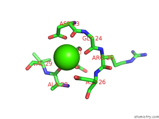
Mono view
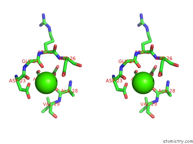
Stereo pair view
Mono view
Stereo pair view
A full contact list of Calcium with other atoms in the Ca binding
site number 1 of Placopecten Striated Muscle Myosin II within 5.0Å range:
|
Reference:
Y.Yang,
S.Gourinath,
M.Kovacs,
L.Nyitray,
R.Reutzel,
D.M.Himmel,
E.O'neall-Hennessey,
L.Reshetnikova,
A.G.Szent-Gyorgyi,
J.H.Brown,
C.Cohen.
Rigor-Like Structures From Muscle Myosins Reveal Key Mechanical Elements in the Transduction Pathways of This Allosteric Motor. Structure V. 15 553 2007.
ISSN: ISSN 0969-2126
PubMed: 17502101
DOI: 10.1016/J.STR.2007.03.010
Page generated: Tue Jul 8 05:19:31 2025
ISSN: ISSN 0969-2126
PubMed: 17502101
DOI: 10.1016/J.STR.2007.03.010
Last articles
Mg in 2UU7Mg in 2UAG
Mg in 2UKD
Mg in 2SHK
Mg in 2TPS
Mg in 2TRT
Mg in 2TRA
Mg in 2RMK
Mg in 2RUS
Mg in 2TCT