Calcium »
PDB 2eex-2f3z »
2exk »
Calcium in PDB 2exk: Structure of the FAMILY43 Beta-Xylosidase E187G From Geobacillus Stearothermophilus in Complex with Xylobiose
Enzymatic activity of Structure of the FAMILY43 Beta-Xylosidase E187G From Geobacillus Stearothermophilus in Complex with Xylobiose
All present enzymatic activity of Structure of the FAMILY43 Beta-Xylosidase E187G From Geobacillus Stearothermophilus in Complex with Xylobiose:
3.2.1.37;
3.2.1.37;
Protein crystallography data
The structure of Structure of the FAMILY43 Beta-Xylosidase E187G From Geobacillus Stearothermophilus in Complex with Xylobiose, PDB code: 2exk
was solved by
C.Brux,
K.Niefind,
D.Shallom-Shezifi,
Y.Shoham,
D.Schomburg,
with X-Ray Crystallography technique. A brief refinement statistics is given in the table below:
| Resolution Low / High (Å) | 2.23 / 2.20 |
| Space group | P 43 21 2 |
| Cell size a, b, c (Å), α, β, γ (°) | 139.870, 139.870, 232.025, 90.00, 90.00, 90.00 |
| R / Rfree (%) | 18.5 / 23.8 |
Calcium Binding Sites:
The binding sites of Calcium atom in the Structure of the FAMILY43 Beta-Xylosidase E187G From Geobacillus Stearothermophilus in Complex with Xylobiose
(pdb code 2exk). This binding sites where shown within
5.0 Angstroms radius around Calcium atom.
In total 4 binding sites of Calcium where determined in the Structure of the FAMILY43 Beta-Xylosidase E187G From Geobacillus Stearothermophilus in Complex with Xylobiose, PDB code: 2exk:
Jump to Calcium binding site number: 1; 2; 3; 4;
In total 4 binding sites of Calcium where determined in the Structure of the FAMILY43 Beta-Xylosidase E187G From Geobacillus Stearothermophilus in Complex with Xylobiose, PDB code: 2exk:
Jump to Calcium binding site number: 1; 2; 3; 4;
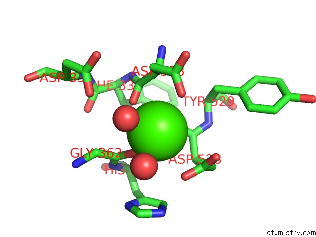
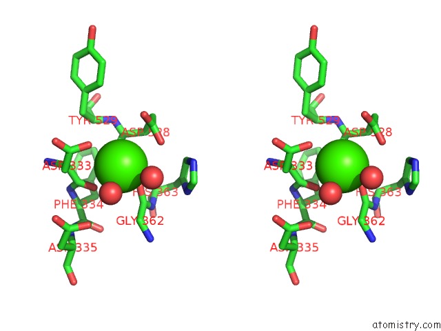
Calcium binding site 1 out of 4 in 2exk
Go back to
Calcium binding site 1 out
of 4 in the Structure of the FAMILY43 Beta-Xylosidase E187G From Geobacillus Stearothermophilus in Complex with Xylobiose
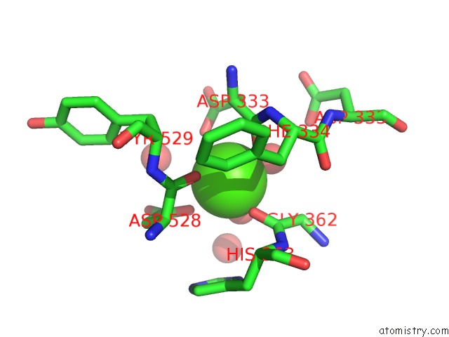
Mono view
Stereo pair view
Mono view
Stereo pair view
A full contact list of Calcium with other atoms in the Ca binding
site number 1 of Structure of the FAMILY43 Beta-Xylosidase E187G From Geobacillus Stearothermophilus in Complex with Xylobiose within 5.0Å range:
|
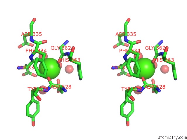
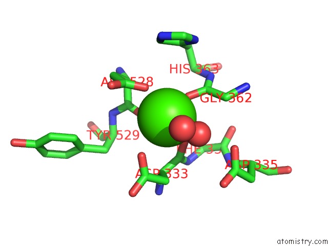
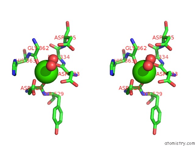
Calcium binding site 2 out of 4 in 2exk
Go back to
Calcium binding site 2 out
of 4 in the Structure of the FAMILY43 Beta-Xylosidase E187G From Geobacillus Stearothermophilus in Complex with Xylobiose
Mono view
Stereo pair view
Mono view
Stereo pair view
A full contact list of Calcium with other atoms in the Ca binding
site number 2 of Structure of the FAMILY43 Beta-Xylosidase E187G From Geobacillus Stearothermophilus in Complex with Xylobiose within 5.0Å range:
|
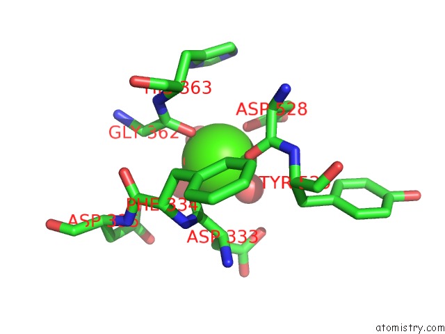
Calcium binding site 3 out of 4 in 2exk
Go back to
Calcium binding site 3 out
of 4 in the Structure of the FAMILY43 Beta-Xylosidase E187G From Geobacillus Stearothermophilus in Complex with Xylobiose
Mono view
Stereo pair view
Mono view
Stereo pair view
A full contact list of Calcium with other atoms in the Ca binding
site number 3 of Structure of the FAMILY43 Beta-Xylosidase E187G From Geobacillus Stearothermophilus in Complex with Xylobiose within 5.0Å range:
|
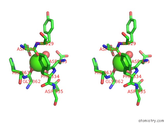
Calcium binding site 4 out of 4 in 2exk
Go back to
Calcium binding site 4 out
of 4 in the Structure of the FAMILY43 Beta-Xylosidase E187G From Geobacillus Stearothermophilus in Complex with Xylobiose
Mono view
Stereo pair view
Mono view
Stereo pair view
A full contact list of Calcium with other atoms in the Ca binding
site number 4 of Structure of the FAMILY43 Beta-Xylosidase E187G From Geobacillus Stearothermophilus in Complex with Xylobiose within 5.0Å range:
|
Reference:
C.Brux,
A.Ben-David,
D.Shallom-Shezifi,
M.Leon,
K.Niefind,
G.Shoham,
Y.Shoham,
D.Schomburg.
The Structure of An Inverting GH43 Beta-Xylosidase From Geobacillus Stearothermophilus with Its Substrate Reveals the Role of the Three Catalytic Residues. J.Mol.Biol. V. 359 97 2006.
ISSN: ISSN 0022-2836
PubMed: 16631196
DOI: 10.1016/J.JMB.2006.03.005
Page generated: Tue Jul 8 05:25:33 2025
ISSN: ISSN 0022-2836
PubMed: 16631196
DOI: 10.1016/J.JMB.2006.03.005
Last articles
K in 5L9WK in 5L9D
K in 5L88
K in 5KSD
K in 5KSE
K in 5K09
K in 5KOE
K in 5KMT
K in 5KIL
K in 5KIK