Calcium »
PDB 2fod-2g81 »
2fpw »
Calcium in PDB 2fpw: Crystal Structure of the N-Terminal Domain of E.Coli Hisb- Phosphoaspartate Intermediate.
Enzymatic activity of Crystal Structure of the N-Terminal Domain of E.Coli Hisb- Phosphoaspartate Intermediate.
All present enzymatic activity of Crystal Structure of the N-Terminal Domain of E.Coli Hisb- Phosphoaspartate Intermediate.:
3.1.3.15;
3.1.3.15;
Protein crystallography data
The structure of Crystal Structure of the N-Terminal Domain of E.Coli Hisb- Phosphoaspartate Intermediate., PDB code: 2fpw
was solved by
E.S.Rangarajan,
M.Cygler,
A.Matte,
Montreal-Kingston Bacterialstructural Genomics Initiative (Bsgi),
with X-Ray Crystallography technique. A brief refinement statistics is given in the table below:
| Resolution Low / High (Å) | 50.00 / 1.75 |
| Space group | C 2 2 21 |
| Cell size a, b, c (Å), α, β, γ (°) | 52.983, 132.408, 105.685, 90.00, 90.00, 90.00 |
| R / Rfree (%) | 17.8 / 21.1 |
Other elements in 2fpw:
The structure of Crystal Structure of the N-Terminal Domain of E.Coli Hisb- Phosphoaspartate Intermediate. also contains other interesting chemical elements:
| Zinc | (Zn) | 2 atoms |
Calcium Binding Sites:
The binding sites of Calcium atom in the Crystal Structure of the N-Terminal Domain of E.Coli Hisb- Phosphoaspartate Intermediate.
(pdb code 2fpw). This binding sites where shown within
5.0 Angstroms radius around Calcium atom.
In total 3 binding sites of Calcium where determined in the Crystal Structure of the N-Terminal Domain of E.Coli Hisb- Phosphoaspartate Intermediate., PDB code: 2fpw:
Jump to Calcium binding site number: 1; 2; 3;
In total 3 binding sites of Calcium where determined in the Crystal Structure of the N-Terminal Domain of E.Coli Hisb- Phosphoaspartate Intermediate., PDB code: 2fpw:
Jump to Calcium binding site number: 1; 2; 3;
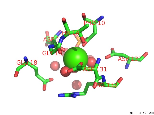
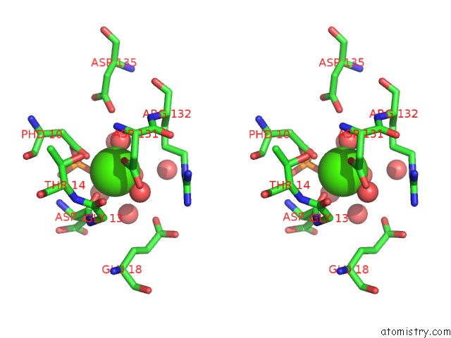
Calcium binding site 1 out of 3 in 2fpw
Go back to
Calcium binding site 1 out
of 3 in the Crystal Structure of the N-Terminal Domain of E.Coli Hisb- Phosphoaspartate Intermediate.
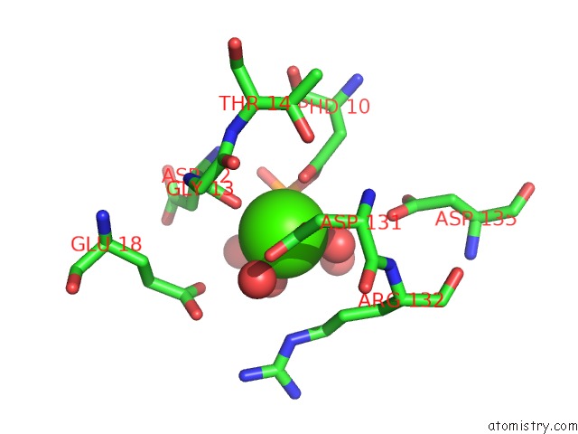
Mono view
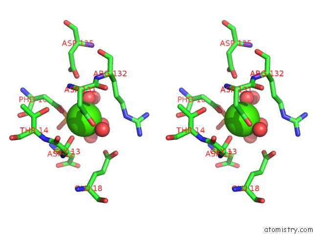
Stereo pair view
Mono view
Stereo pair view
A full contact list of Calcium with other atoms in the Ca binding
site number 1 of Crystal Structure of the N-Terminal Domain of E.Coli Hisb- Phosphoaspartate Intermediate. within 5.0Å range:
|
Calcium binding site 2 out of 3 in 2fpw
Go back to
Calcium binding site 2 out
of 3 in the Crystal Structure of the N-Terminal Domain of E.Coli Hisb- Phosphoaspartate Intermediate.
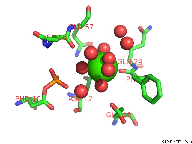
Mono view
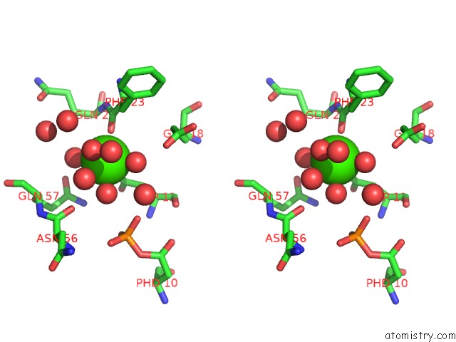
Stereo pair view
Mono view
Stereo pair view
A full contact list of Calcium with other atoms in the Ca binding
site number 2 of Crystal Structure of the N-Terminal Domain of E.Coli Hisb- Phosphoaspartate Intermediate. within 5.0Å range:
|
Calcium binding site 3 out of 3 in 2fpw
Go back to
Calcium binding site 3 out
of 3 in the Crystal Structure of the N-Terminal Domain of E.Coli Hisb- Phosphoaspartate Intermediate.
Mono view
Stereo pair view
Mono view
Stereo pair view
A full contact list of Calcium with other atoms in the Ca binding
site number 3 of Crystal Structure of the N-Terminal Domain of E.Coli Hisb- Phosphoaspartate Intermediate. within 5.0Å range:
|
Reference:
E.S.Rangarajan,
A.Proteau,
J.Wagner,
M.N.Hung,
A.Matte,
M.Cygler.
Structural Snapshots of Escherichia Coli Histidinol Phosphate Phosphatase Along the Reaction Pathway. J.Biol.Chem. V. 281 37930 2006.
ISSN: ISSN 0021-9258
PubMed: 16966333
DOI: 10.1074/JBC.M604916200
Page generated: Tue Jul 8 05:37:25 2025
ISSN: ISSN 0021-9258
PubMed: 16966333
DOI: 10.1074/JBC.M604916200
Last articles
Mn in 2G8IMn in 2GA2
Mn in 2G73
Mn in 2G74
Mn in 2G38
Mn in 2FYD
Mn in 2G4I
Mn in 2FYB
Mn in 2FV2
Mn in 2FYA