Calcium »
PDB 2g8e-2gsm »
2gsm »
Calcium in PDB 2gsm: Catalytic Core (Subunits I and II) of Cytochrome C Oxidase From Rhodobacter Sphaeroides
Enzymatic activity of Catalytic Core (Subunits I and II) of Cytochrome C Oxidase From Rhodobacter Sphaeroides
All present enzymatic activity of Catalytic Core (Subunits I and II) of Cytochrome C Oxidase From Rhodobacter Sphaeroides:
1.9.3.1;
1.9.3.1;
Protein crystallography data
The structure of Catalytic Core (Subunits I and II) of Cytochrome C Oxidase From Rhodobacter Sphaeroides, PDB code: 2gsm
was solved by
L.Qin,
C.Hiser,
A.Mulichak,
R.M.Garavito,
S.Ferguson-Miller,
with X-Ray Crystallography technique. A brief refinement statistics is given in the table below:
| Resolution Low / High (Å) | 20.00 / 2.00 |
| Space group | P 21 21 21 |
| Cell size a, b, c (Å), α, β, γ (°) | 125.020, 131.639, 176.802, 90.00, 90.00, 90.00 |
| R / Rfree (%) | 21.4 / 23.2 |
Other elements in 2gsm:
The structure of Catalytic Core (Subunits I and II) of Cytochrome C Oxidase From Rhodobacter Sphaeroides also contains other interesting chemical elements:
| Magnesium | (Mg) | 2 atoms |
| Cadmium | (Cd) | 4 atoms |
| Iron | (Fe) | 4 atoms |
| Copper | (Cu) | 6 atoms |
Calcium Binding Sites:
The binding sites of Calcium atom in the Catalytic Core (Subunits I and II) of Cytochrome C Oxidase From Rhodobacter Sphaeroides
(pdb code 2gsm). This binding sites where shown within
5.0 Angstroms radius around Calcium atom.
In total 2 binding sites of Calcium where determined in the Catalytic Core (Subunits I and II) of Cytochrome C Oxidase From Rhodobacter Sphaeroides, PDB code: 2gsm:
Jump to Calcium binding site number: 1; 2;
In total 2 binding sites of Calcium where determined in the Catalytic Core (Subunits I and II) of Cytochrome C Oxidase From Rhodobacter Sphaeroides, PDB code: 2gsm:
Jump to Calcium binding site number: 1; 2;
Calcium binding site 1 out of 2 in 2gsm
Go back to
Calcium binding site 1 out
of 2 in the Catalytic Core (Subunits I and II) of Cytochrome C Oxidase From Rhodobacter Sphaeroides
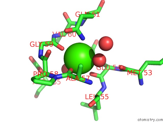
Mono view
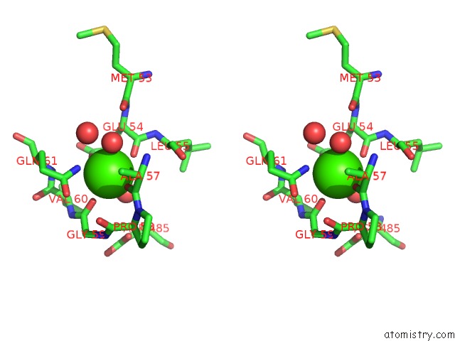
Stereo pair view
Mono view
Stereo pair view
A full contact list of Calcium with other atoms in the Ca binding
site number 1 of Catalytic Core (Subunits I and II) of Cytochrome C Oxidase From Rhodobacter Sphaeroides within 5.0Å range:
|
Calcium binding site 2 out of 2 in 2gsm
Go back to
Calcium binding site 2 out
of 2 in the Catalytic Core (Subunits I and II) of Cytochrome C Oxidase From Rhodobacter Sphaeroides
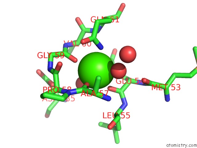
Mono view

Stereo pair view
Mono view
Stereo pair view
A full contact list of Calcium with other atoms in the Ca binding
site number 2 of Catalytic Core (Subunits I and II) of Cytochrome C Oxidase From Rhodobacter Sphaeroides within 5.0Å range:
|
Reference:
L.Qin,
C.Hiser,
A.Mulichak,
R.M.Garavito,
S.Ferguson-Miller.
Identification of Conserved Lipid/Detergent-Binding Sites in A High-Resolution Structure of the Membrane Protein Cytochrome C Oxidase. Proc.Natl.Acad.Sci.Usa V. 103 16117 2006.
ISSN: ISSN 0027-8424
PubMed: 17050688
DOI: 10.1073/PNAS.0606149103
Page generated: Tue Jul 8 05:50:21 2025
ISSN: ISSN 0027-8424
PubMed: 17050688
DOI: 10.1073/PNAS.0606149103
Last articles
I in 1UO5I in 1UJK
I in 1UO4
I in 1T1F
I in 1SN5
I in 1UHI
I in 1UE2
I in 1U65
I in 1THA
I in 1TUK