Calcium »
PDB 2gsp-2hib »
2gwk »
Calcium in PDB 2gwk: Spvb Adp-Ribosylated Actin: Orthorhombic Crystal Form
Protein crystallography data
The structure of Spvb Adp-Ribosylated Actin: Orthorhombic Crystal Form, PDB code: 2gwk
was solved by
C.E.Stebbins,
S.M.Margarit,
with X-Ray Crystallography technique. A brief refinement statistics is given in the table below:
| Resolution Low / High (Å) | 32.00 / 2.00 |
| Space group | P 21 21 21 |
| Cell size a, b, c (Å), α, β, γ (°) | 100.385, 102.348, 123.803, 90.00, 90.00, 90.00 |
| R / Rfree (%) | 17.1 / 21.1 |
Calcium Binding Sites:
The binding sites of Calcium atom in the Spvb Adp-Ribosylated Actin: Orthorhombic Crystal Form
(pdb code 2gwk). This binding sites where shown within
5.0 Angstroms radius around Calcium atom.
In total 2 binding sites of Calcium where determined in the Spvb Adp-Ribosylated Actin: Orthorhombic Crystal Form, PDB code: 2gwk:
Jump to Calcium binding site number: 1; 2;
In total 2 binding sites of Calcium where determined in the Spvb Adp-Ribosylated Actin: Orthorhombic Crystal Form, PDB code: 2gwk:
Jump to Calcium binding site number: 1; 2;
Calcium binding site 1 out of 2 in 2gwk
Go back to
Calcium binding site 1 out
of 2 in the Spvb Adp-Ribosylated Actin: Orthorhombic Crystal Form
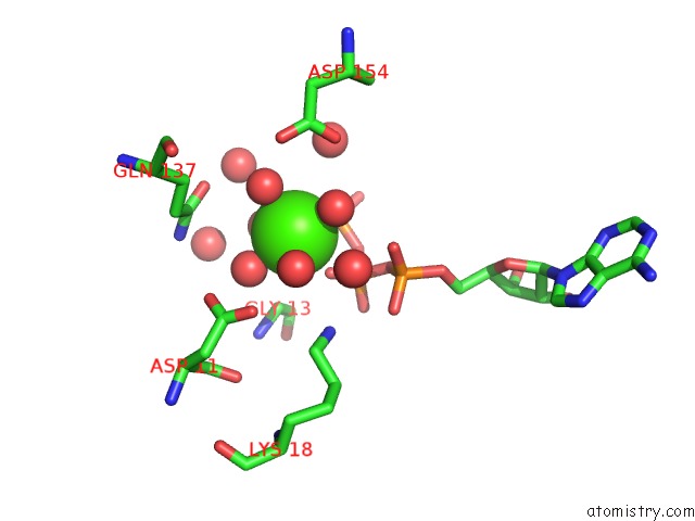
Mono view
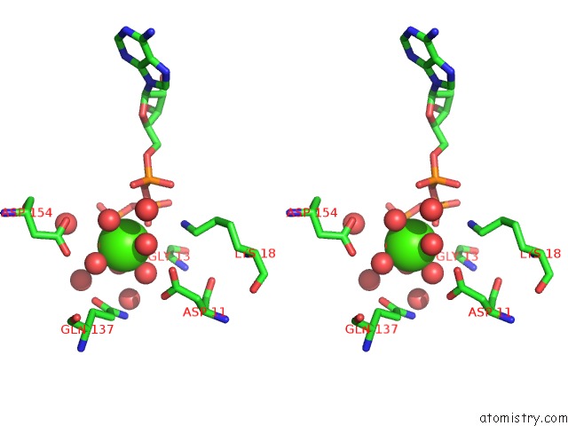
Stereo pair view
Mono view
Stereo pair view
A full contact list of Calcium with other atoms in the Ca binding
site number 1 of Spvb Adp-Ribosylated Actin: Orthorhombic Crystal Form within 5.0Å range:
|
Calcium binding site 2 out of 2 in 2gwk
Go back to
Calcium binding site 2 out
of 2 in the Spvb Adp-Ribosylated Actin: Orthorhombic Crystal Form
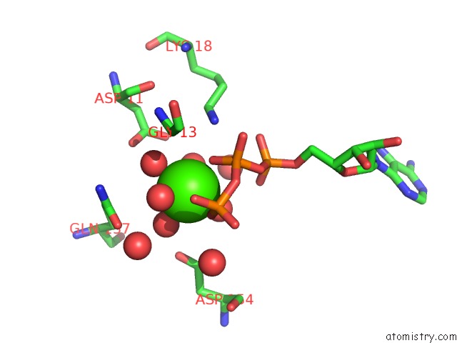
Mono view
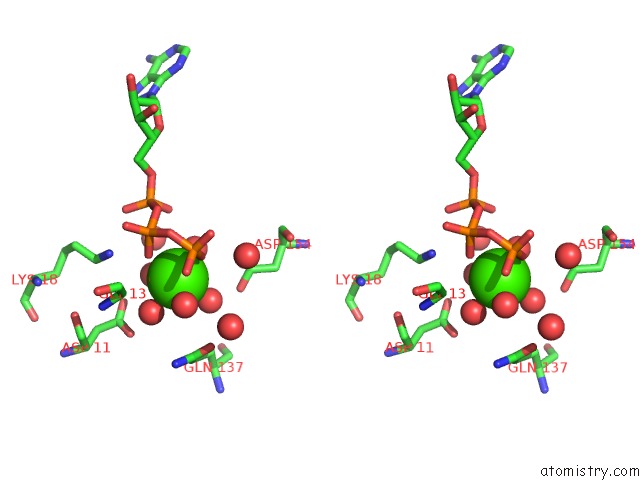
Stereo pair view
Mono view
Stereo pair view
A full contact list of Calcium with other atoms in the Ca binding
site number 2 of Spvb Adp-Ribosylated Actin: Orthorhombic Crystal Form within 5.0Å range:
|
Reference:
S.M.Margarit,
W.Davidson,
L.Frego,
C.E.Stebbins.
A Steric Antagonism of Actin Polymerization By A Salmonella Virulence Protein. Structure V. 14 1219 2006.
ISSN: ISSN 0969-2126
PubMed: 16905096
DOI: 10.1016/J.STR.2006.05.022
Page generated: Tue Jul 8 05:52:21 2025
ISSN: ISSN 0969-2126
PubMed: 16905096
DOI: 10.1016/J.STR.2006.05.022
Last articles
I in 4TQDI in 4TMD
I in 4S3Q
I in 4TJV
I in 4S22
I in 4S2H
I in 4QX5
I in 4S2G
I in 4S2F
I in 4RX1