Calcium »
PDB 2gsp-2hib »
2gy7 »
Calcium in PDB 2gy7: Angiopoietin-2/TIE2 Complex Crystal Structure
Enzymatic activity of Angiopoietin-2/TIE2 Complex Crystal Structure
All present enzymatic activity of Angiopoietin-2/TIE2 Complex Crystal Structure:
2.7.10.1;
2.7.10.1;
Protein crystallography data
The structure of Angiopoietin-2/TIE2 Complex Crystal Structure, PDB code: 2gy7
was solved by
W.A.Barton,
D.B.Nikolov,
with X-Ray Crystallography technique. A brief refinement statistics is given in the table below:
| Resolution Low / High (Å) | 8.00 / 3.70 |
| Space group | P 41 21 2 |
| Cell size a, b, c (Å), α, β, γ (°) | 165.636, 165.636, 115.310, 90.00, 90.00, 90.00 |
| R / Rfree (%) | 24 / 31.7 |
Calcium Binding Sites:
The binding sites of Calcium atom in the Angiopoietin-2/TIE2 Complex Crystal Structure
(pdb code 2gy7). This binding sites where shown within
5.0 Angstroms radius around Calcium atom.
In total only one binding site of Calcium was determined in the Angiopoietin-2/TIE2 Complex Crystal Structure, PDB code: 2gy7:
In total only one binding site of Calcium was determined in the Angiopoietin-2/TIE2 Complex Crystal Structure, PDB code: 2gy7:
Calcium binding site 1 out of 1 in 2gy7
Go back to
Calcium binding site 1 out
of 1 in the Angiopoietin-2/TIE2 Complex Crystal Structure

Mono view
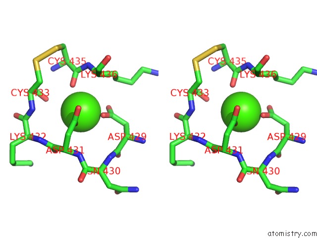
Stereo pair view
Mono view
Stereo pair view
A full contact list of Calcium with other atoms in the Ca binding
site number 1 of Angiopoietin-2/TIE2 Complex Crystal Structure within 5.0Å range:
|
Reference:
W.A.Barton,
D.Tzvetkova-Robev,
E.P.Miranda,
M.V.Kolev,
K.R.Rajashankar,
J.P.Himanen,
D.B.Nikolov.
Crystal Structures of the TIE2 Receptor Ectodomain and the Angiopoietin-2-TIE2 Complex. Nat.Struct.Mol.Biol. V. 13 524 2006.
ISSN: ISSN 1545-9993
PubMed: 16732286
DOI: 10.1038/NSMB1101
Page generated: Tue Jul 8 05:52:45 2025
ISSN: ISSN 1545-9993
PubMed: 16732286
DOI: 10.1038/NSMB1101
Last articles
I in 5BJOI in 5AV3
I in 5AUR
I in 5A7H
I in 5AX3
I in 5AOJ
I in 4ZU2
I in 5AKX
I in 5A3D
I in 4ZV0