Calcium »
PDB 2gsp-2hib »
2h0l »
Calcium in PDB 2h0l: Crystal Structure of A Mutant of Rat Annexin A5
Protein crystallography data
The structure of Crystal Structure of A Mutant of Rat Annexin A5, PDB code: 2h0l
was solved by
B.Langlois D'estaintot,
B.Gallois,
T.Granier,
B.Tessier,
A.Brisson,
with X-Ray Crystallography technique. A brief refinement statistics is given in the table below:
| Resolution Low / High (Å) | 32.76 / 2.59 |
| Space group | P 21 21 2 |
| Cell size a, b, c (Å), α, β, γ (°) | 77.918, 78.959, 60.458, 90.00, 90.00, 90.00 |
| R / Rfree (%) | 18.7 / 27.8 |
Calcium Binding Sites:
The binding sites of Calcium atom in the Crystal Structure of A Mutant of Rat Annexin A5
(pdb code 2h0l). This binding sites where shown within
5.0 Angstroms radius around Calcium atom.
In total 3 binding sites of Calcium where determined in the Crystal Structure of A Mutant of Rat Annexin A5, PDB code: 2h0l:
Jump to Calcium binding site number: 1; 2; 3;
In total 3 binding sites of Calcium where determined in the Crystal Structure of A Mutant of Rat Annexin A5, PDB code: 2h0l:
Jump to Calcium binding site number: 1; 2; 3;
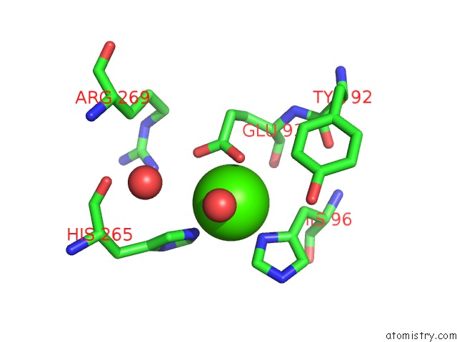
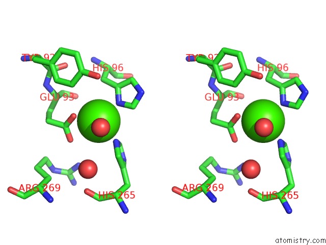
Calcium binding site 1 out of 3 in 2h0l
Go back to
Calcium binding site 1 out
of 3 in the Crystal Structure of A Mutant of Rat Annexin A5
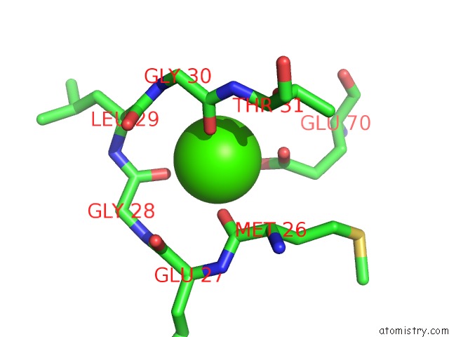
Mono view
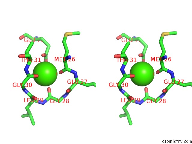
Stereo pair view
Mono view
Stereo pair view
A full contact list of Calcium with other atoms in the Ca binding
site number 1 of Crystal Structure of A Mutant of Rat Annexin A5 within 5.0Å range:
|
Calcium binding site 2 out of 3 in 2h0l
Go back to
Calcium binding site 2 out
of 3 in the Crystal Structure of A Mutant of Rat Annexin A5
Mono view
Stereo pair view
Mono view
Stereo pair view
A full contact list of Calcium with other atoms in the Ca binding
site number 2 of Crystal Structure of A Mutant of Rat Annexin A5 within 5.0Å range:
|
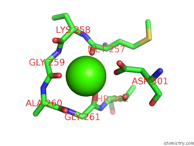
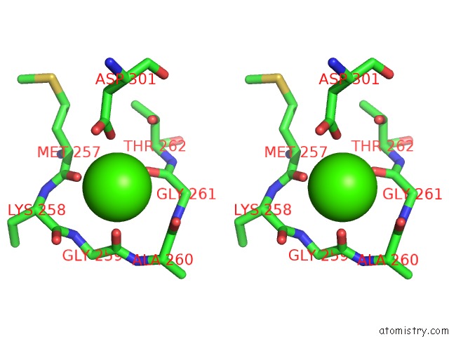
Calcium binding site 3 out of 3 in 2h0l
Go back to
Calcium binding site 3 out
of 3 in the Crystal Structure of A Mutant of Rat Annexin A5
Mono view
Stereo pair view
Mono view
Stereo pair view
A full contact list of Calcium with other atoms in the Ca binding
site number 3 of Crystal Structure of A Mutant of Rat Annexin A5 within 5.0Å range:
|
Reference:
A.Brisson,
T.Granier,
B.Langlois D'estaintot,
B.Gallois,
B.Tessier.
Identification of the Residues Involved in the Formation of Annexin V Trimers Within 2D and 3D Crystals To Be Published.
Page generated: Tue Jul 8 05:53:50 2025
Last articles
I in 4TQDI in 4TMD
I in 4S3Q
I in 4TJV
I in 4S22
I in 4S2H
I in 4QX5
I in 4S2G
I in 4S2F
I in 4RX1