Calcium »
PDB 2hih-2i52 »
2hpc »
Calcium in PDB 2hpc: Crystal Structure of Fragment D From Human Fibrinogen Complexed with Gly-Pro-Arg-Pro-Amide.
Protein crystallography data
The structure of Crystal Structure of Fragment D From Human Fibrinogen Complexed with Gly-Pro-Arg-Pro-Amide., PDB code: 2hpc
was solved by
R.F.Doolittle,
J.M.Kollman,
A.Chen,
L.Pandi,
with X-Ray Crystallography technique. A brief refinement statistics is given in the table below:
| Resolution Low / High (Å) | 30.00 / 2.90 |
| Space group | P 1 2 1 |
| Cell size a, b, c (Å), α, β, γ (°) | 81.677, 46.069, 429.901, 90.00, 89.99, 90.00 |
| R / Rfree (%) | 27.5 / 36 |
Calcium Binding Sites:
The binding sites of Calcium atom in the Crystal Structure of Fragment D From Human Fibrinogen Complexed with Gly-Pro-Arg-Pro-Amide.
(pdb code 2hpc). This binding sites where shown within
5.0 Angstroms radius around Calcium atom.
In total 10 binding sites of Calcium where determined in the Crystal Structure of Fragment D From Human Fibrinogen Complexed with Gly-Pro-Arg-Pro-Amide., PDB code: 2hpc:
Jump to Calcium binding site number: 1; 2; 3; 4; 5; 6; 7; 8; 9; 10;
In total 10 binding sites of Calcium where determined in the Crystal Structure of Fragment D From Human Fibrinogen Complexed with Gly-Pro-Arg-Pro-Amide., PDB code: 2hpc:
Jump to Calcium binding site number: 1; 2; 3; 4; 5; 6; 7; 8; 9; 10;
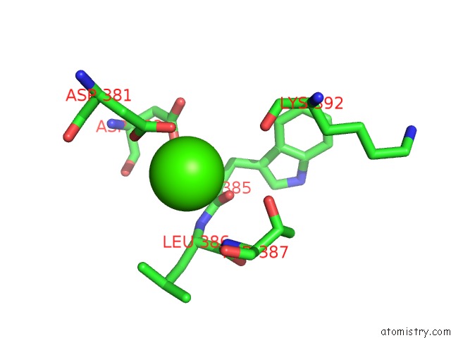
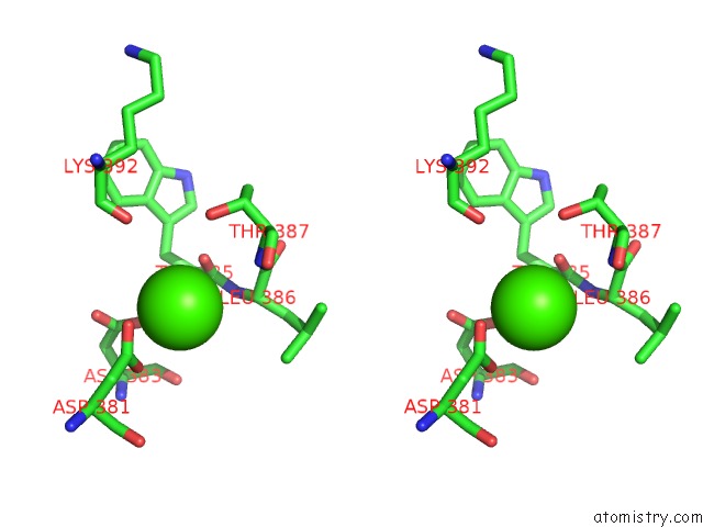
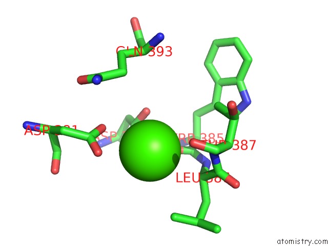
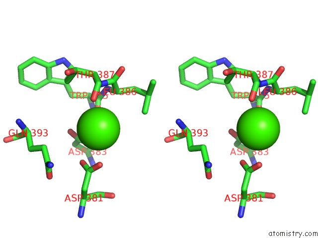
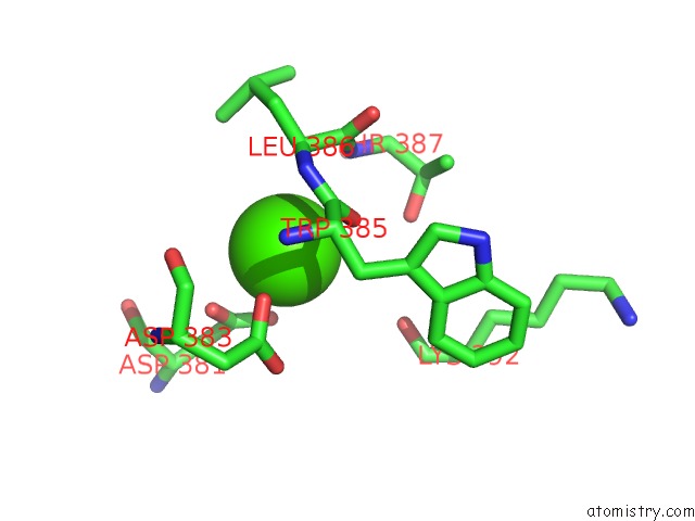
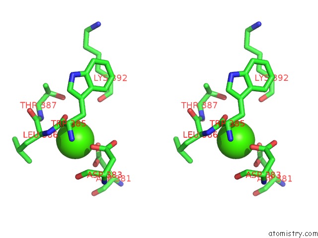
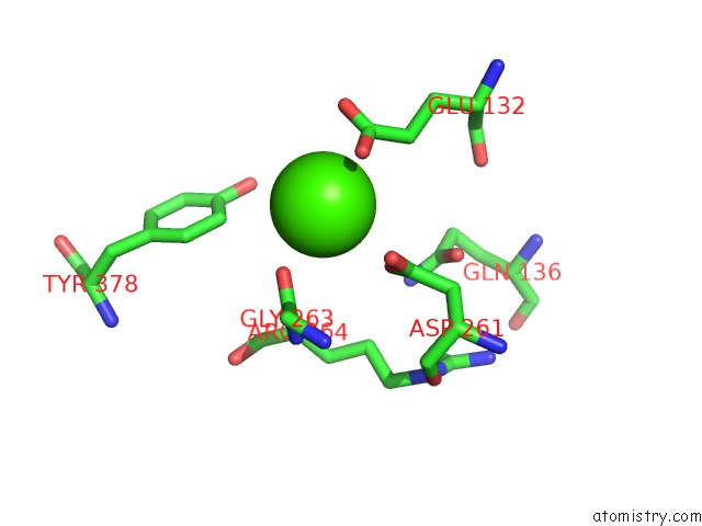
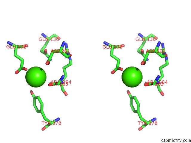
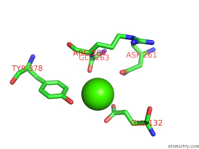
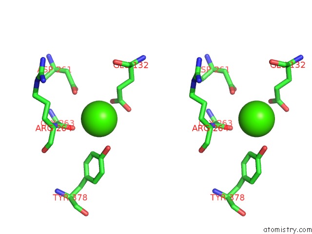
Calcium binding site 1 out of 10 in 2hpc
Go back to
Calcium binding site 1 out
of 10 in the Crystal Structure of Fragment D From Human Fibrinogen Complexed with Gly-Pro-Arg-Pro-Amide.
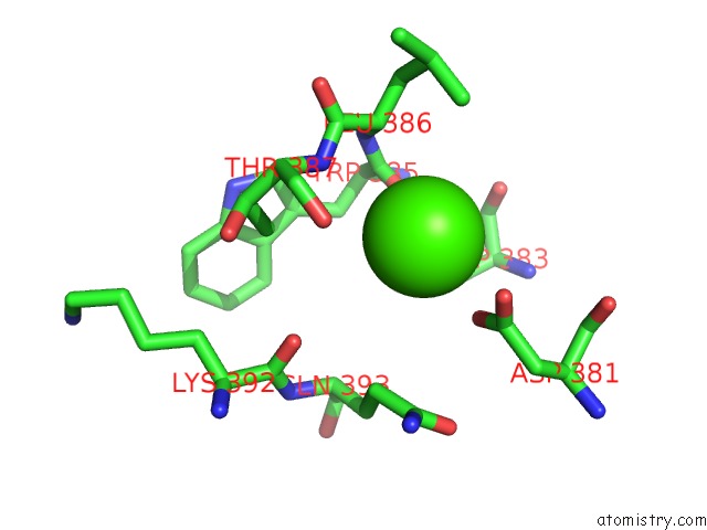
Mono view
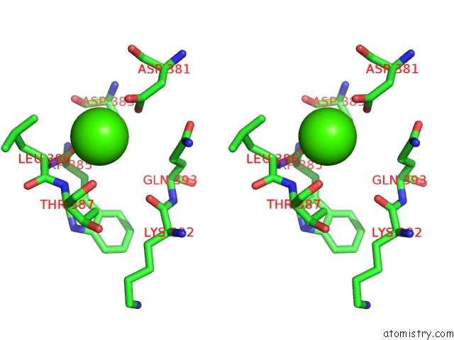
Stereo pair view
Mono view
Stereo pair view
A full contact list of Calcium with other atoms in the Ca binding
site number 1 of Crystal Structure of Fragment D From Human Fibrinogen Complexed with Gly-Pro-Arg-Pro-Amide. within 5.0Å range:
|
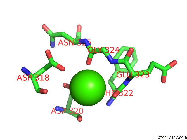
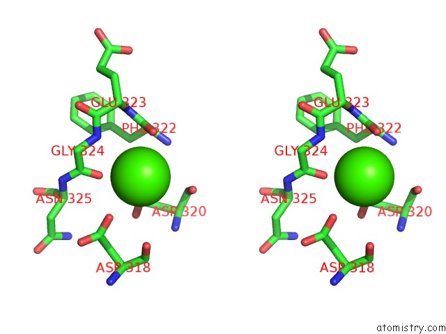
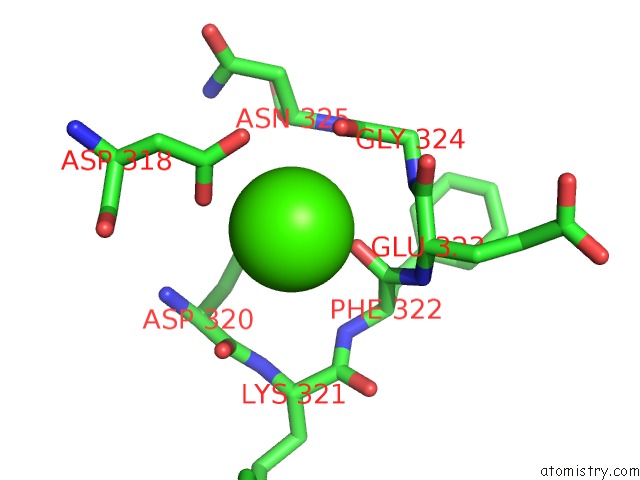
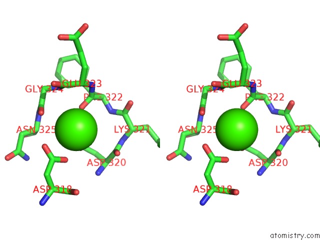
Calcium binding site 2 out of 10 in 2hpc
Go back to
Calcium binding site 2 out
of 10 in the Crystal Structure of Fragment D From Human Fibrinogen Complexed with Gly-Pro-Arg-Pro-Amide.
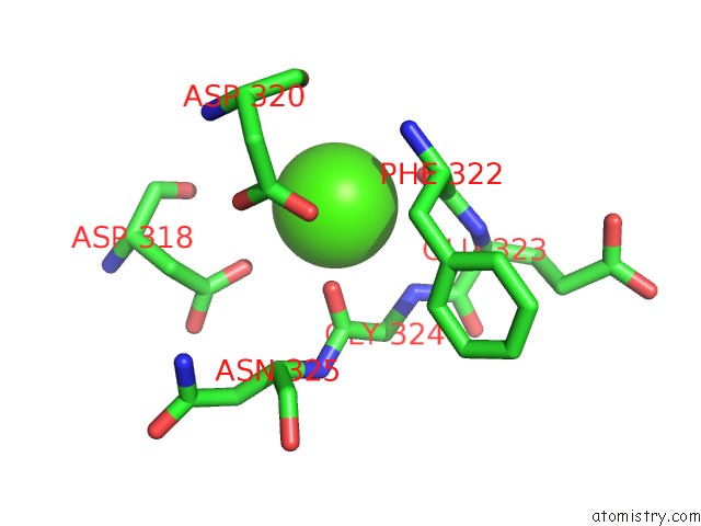
Mono view
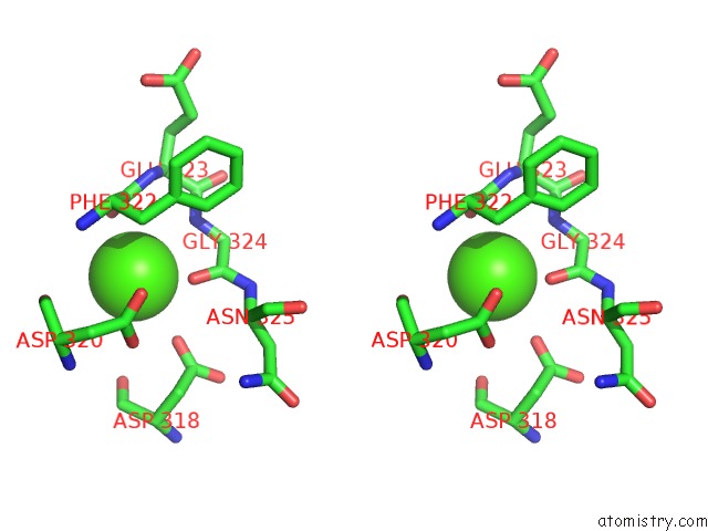
Stereo pair view
Mono view
Stereo pair view
A full contact list of Calcium with other atoms in the Ca binding
site number 2 of Crystal Structure of Fragment D From Human Fibrinogen Complexed with Gly-Pro-Arg-Pro-Amide. within 5.0Å range:
|
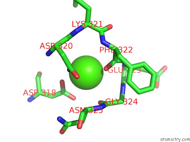
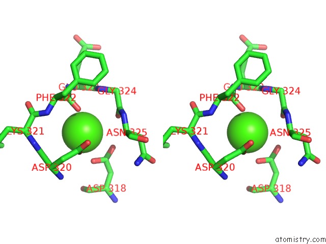
Calcium binding site 3 out of 10 in 2hpc
Go back to
Calcium binding site 3 out
of 10 in the Crystal Structure of Fragment D From Human Fibrinogen Complexed with Gly-Pro-Arg-Pro-Amide.
Mono view
Stereo pair view
Mono view
Stereo pair view
A full contact list of Calcium with other atoms in the Ca binding
site number 3 of Crystal Structure of Fragment D From Human Fibrinogen Complexed with Gly-Pro-Arg-Pro-Amide. within 5.0Å range:
|
Calcium binding site 4 out of 10 in 2hpc
Go back to
Calcium binding site 4 out
of 10 in the Crystal Structure of Fragment D From Human Fibrinogen Complexed with Gly-Pro-Arg-Pro-Amide.
Mono view
Stereo pair view
Mono view
Stereo pair view
A full contact list of Calcium with other atoms in the Ca binding
site number 4 of Crystal Structure of Fragment D From Human Fibrinogen Complexed with Gly-Pro-Arg-Pro-Amide. within 5.0Å range:
|
Calcium binding site 5 out of 10 in 2hpc
Go back to
Calcium binding site 5 out
of 10 in the Crystal Structure of Fragment D From Human Fibrinogen Complexed with Gly-Pro-Arg-Pro-Amide.
Mono view
Stereo pair view
Mono view
Stereo pair view
A full contact list of Calcium with other atoms in the Ca binding
site number 5 of Crystal Structure of Fragment D From Human Fibrinogen Complexed with Gly-Pro-Arg-Pro-Amide. within 5.0Å range:
|
Calcium binding site 6 out of 10 in 2hpc
Go back to
Calcium binding site 6 out
of 10 in the Crystal Structure of Fragment D From Human Fibrinogen Complexed with Gly-Pro-Arg-Pro-Amide.
Mono view
Stereo pair view
Mono view
Stereo pair view
A full contact list of Calcium with other atoms in the Ca binding
site number 6 of Crystal Structure of Fragment D From Human Fibrinogen Complexed with Gly-Pro-Arg-Pro-Amide. within 5.0Å range:
|
Calcium binding site 7 out of 10 in 2hpc
Go back to
Calcium binding site 7 out
of 10 in the Crystal Structure of Fragment D From Human Fibrinogen Complexed with Gly-Pro-Arg-Pro-Amide.
Mono view
Stereo pair view
Mono view
Stereo pair view
A full contact list of Calcium with other atoms in the Ca binding
site number 7 of Crystal Structure of Fragment D From Human Fibrinogen Complexed with Gly-Pro-Arg-Pro-Amide. within 5.0Å range:
|
Calcium binding site 8 out of 10 in 2hpc
Go back to
Calcium binding site 8 out
of 10 in the Crystal Structure of Fragment D From Human Fibrinogen Complexed with Gly-Pro-Arg-Pro-Amide.
Mono view
Stereo pair view
Mono view
Stereo pair view
A full contact list of Calcium with other atoms in the Ca binding
site number 8 of Crystal Structure of Fragment D From Human Fibrinogen Complexed with Gly-Pro-Arg-Pro-Amide. within 5.0Å range:
|
Calcium binding site 9 out of 10 in 2hpc
Go back to
Calcium binding site 9 out
of 10 in the Crystal Structure of Fragment D From Human Fibrinogen Complexed with Gly-Pro-Arg-Pro-Amide.
Mono view
Stereo pair view
Mono view
Stereo pair view
A full contact list of Calcium with other atoms in the Ca binding
site number 9 of Crystal Structure of Fragment D From Human Fibrinogen Complexed with Gly-Pro-Arg-Pro-Amide. within 5.0Å range:
|
Calcium binding site 10 out of 10 in 2hpc
Go back to
Calcium binding site 10 out
of 10 in the Crystal Structure of Fragment D From Human Fibrinogen Complexed with Gly-Pro-Arg-Pro-Amide.
Mono view
Stereo pair view
Mono view
Stereo pair view
A full contact list of Calcium with other atoms in the Ca binding
site number 10 of Crystal Structure of Fragment D From Human Fibrinogen Complexed with Gly-Pro-Arg-Pro-Amide. within 5.0Å range:
|
Reference:
R.F.Doolittle,
J.M.Kollman,
A.Chen,
L.Pandi.
N/A N/A.
Page generated: Tue Jul 8 06:02:30 2025
Last articles
Mg in 5ZCQMg in 5ZCP
Mg in 5ZCR
Mg in 5ZCL
Mg in 5ZCH
Mg in 5ZCO
Mg in 5ZCG
Mg in 5ZB0
Mg in 5ZC6
Mg in 5ZC9