Calcium »
PDB 2hih-2i52 »
2i4c »
Calcium in PDB 2i4c: Crystal Structure of Bicarbonate Transport Protein Cmpa From Synechocystis Sp. Pcc 6803 in Complex with Bicarbonate and Calcium
Protein crystallography data
The structure of Crystal Structure of Bicarbonate Transport Protein Cmpa From Synechocystis Sp. Pcc 6803 in Complex with Bicarbonate and Calcium, PDB code: 2i4c
was solved by
N.M.Koropatkin,
T.J.Smith,
H.B.Pakrasi,
with X-Ray Crystallography technique. A brief refinement statistics is given in the table below:
| Resolution Low / High (Å) | 56.82 / 1.70 |
| Space group | P 1 |
| Cell size a, b, c (Å), α, β, γ (°) | 44.240, 49.240, 57.200, 87.40, 80.80, 75.40 |
| R / Rfree (%) | 19.3 / 21.8 |
Calcium Binding Sites:
The binding sites of Calcium atom in the Crystal Structure of Bicarbonate Transport Protein Cmpa From Synechocystis Sp. Pcc 6803 in Complex with Bicarbonate and Calcium
(pdb code 2i4c). This binding sites where shown within
5.0 Angstroms radius around Calcium atom.
In total only one binding site of Calcium was determined in the Crystal Structure of Bicarbonate Transport Protein Cmpa From Synechocystis Sp. Pcc 6803 in Complex with Bicarbonate and Calcium, PDB code: 2i4c:
In total only one binding site of Calcium was determined in the Crystal Structure of Bicarbonate Transport Protein Cmpa From Synechocystis Sp. Pcc 6803 in Complex with Bicarbonate and Calcium, PDB code: 2i4c:
Calcium binding site 1 out of 1 in 2i4c
Go back to
Calcium binding site 1 out
of 1 in the Crystal Structure of Bicarbonate Transport Protein Cmpa From Synechocystis Sp. Pcc 6803 in Complex with Bicarbonate and Calcium
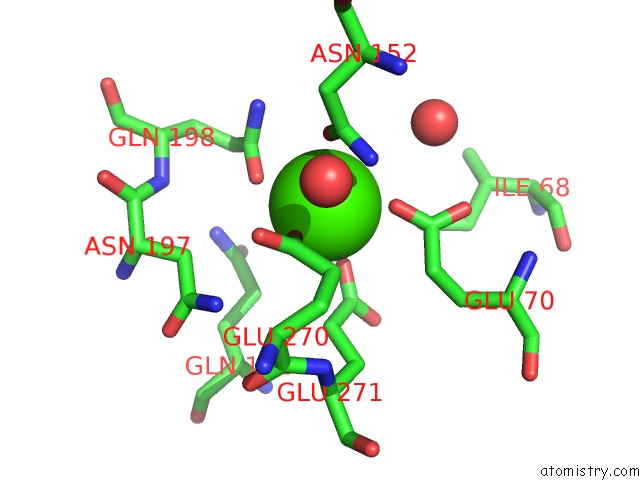
Mono view
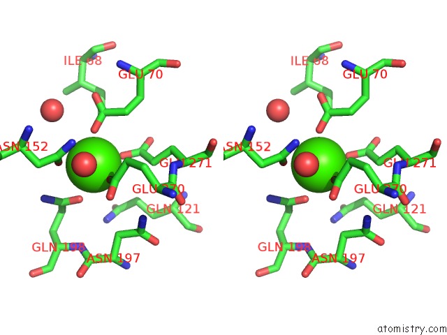
Stereo pair view
Mono view
Stereo pair view
A full contact list of Calcium with other atoms in the Ca binding
site number 1 of Crystal Structure of Bicarbonate Transport Protein Cmpa From Synechocystis Sp. Pcc 6803 in Complex with Bicarbonate and Calcium within 5.0Å range:
|
Reference:
N.M.Koropatkin,
D.W.Koppenaal,
H.B.Pakrasi,
T.J.Smith.
The Structure of A Cyanobacterial Bicarbonate Transport Protein, Cmpa. J.Biol.Chem. V. 282 2606 2007.
ISSN: ISSN 0021-9258
PubMed: 17121816
DOI: 10.1074/JBC.M610222200
Page generated: Tue Jul 8 06:08:41 2025
ISSN: ISSN 0021-9258
PubMed: 17121816
DOI: 10.1074/JBC.M610222200
Last articles
Mg in 6D3QMg in 6D83
Mg in 6D71
Mg in 6D5X
Mg in 6D6R
Mg in 6D6Q
Mg in 6D5W
Mg in 6D5V
Mg in 6D5M
Mg in 6D5K