Calcium »
PDB 2hih-2i52 »
2i52 »
Calcium in PDB 2i52: Crystal Structure of Protein PTO0218 From Picrophilus Torridus, Pfam DUF372
Protein crystallography data
The structure of Crystal Structure of Protein PTO0218 From Picrophilus Torridus, Pfam DUF372, PDB code: 2i52
was solved by
U.A.Ramagopal,
J.Gilmore,
R.Toro,
K.T.Bain,
C.Mckenzie,
C.Reyes,
J.M.Sauder,
S.K.Burley,
S.C.Almo,
New York Sgx Research Center Forstructural Genomics (Nysgxrc),
with X-Ray Crystallography technique. A brief refinement statistics is given in the table below:
| Resolution Low / High (Å) | 40.20 / 2.08 |
| Space group | C 2 2 21 |
| Cell size a, b, c (Å), α, β, γ (°) | 90.764, 143.485, 129.799, 90.00, 90.00, 90.00 |
| R / Rfree (%) | 17.6 / 22.5 |
Other elements in 2i52:
The structure of Crystal Structure of Protein PTO0218 From Picrophilus Torridus, Pfam DUF372 also contains other interesting chemical elements:
| Chlorine | (Cl) | 1 atom |
Calcium Binding Sites:
Pages:
>>> Page 1 <<< Page 2, Binding sites: 11 - 13;Binding sites:
The binding sites of Calcium atom in the Crystal Structure of Protein PTO0218 From Picrophilus Torridus, Pfam DUF372 (pdb code 2i52). This binding sites where shown within 5.0 Angstroms radius around Calcium atom.In total 13 binding sites of Calcium where determined in the Crystal Structure of Protein PTO0218 From Picrophilus Torridus, Pfam DUF372, PDB code: 2i52:
Jump to Calcium binding site number: 1; 2; 3; 4; 5; 6; 7; 8; 9; 10;
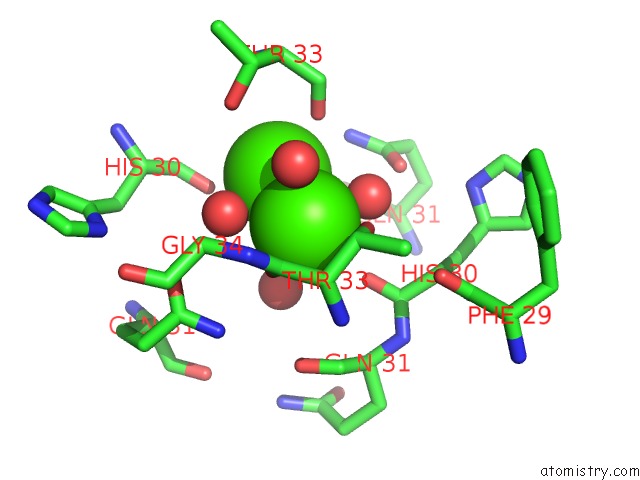
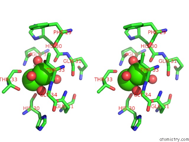
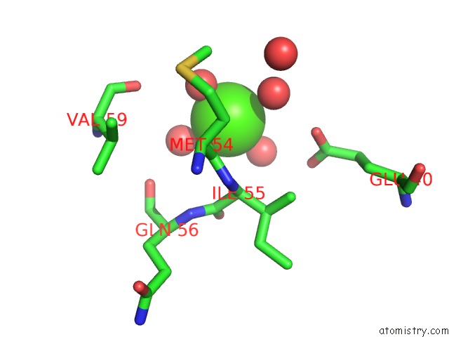
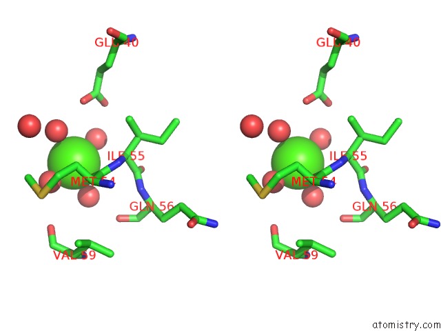
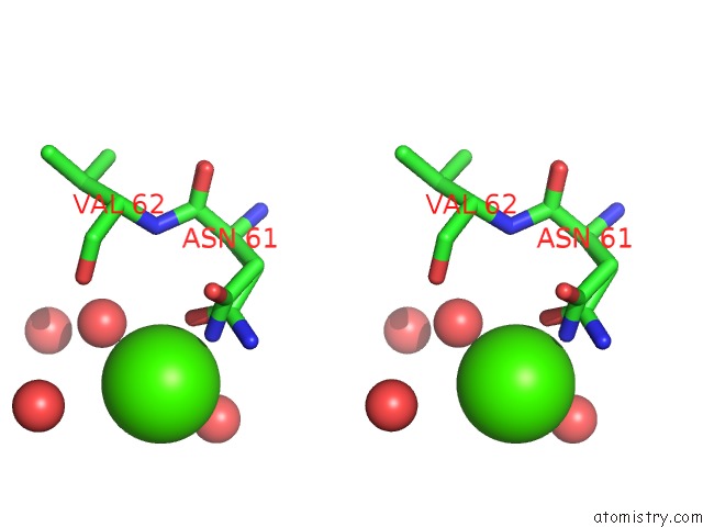
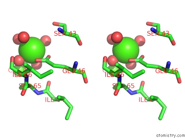
Calcium binding site 1 out of 13 in 2i52
Go back to
Calcium binding site 1 out
of 13 in the Crystal Structure of Protein PTO0218 From Picrophilus Torridus, Pfam DUF372
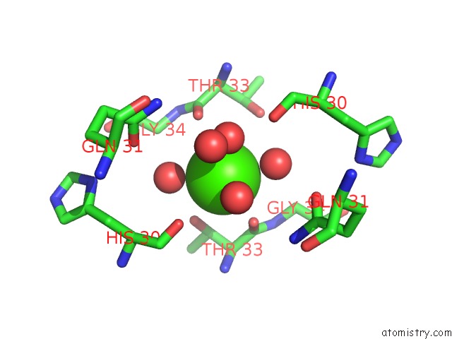
Mono view
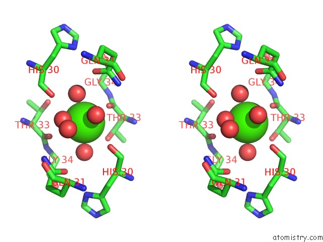
Stereo pair view
Mono view
Stereo pair view
A full contact list of Calcium with other atoms in the Ca binding
site number 1 of Crystal Structure of Protein PTO0218 From Picrophilus Torridus, Pfam DUF372 within 5.0Å range:
|
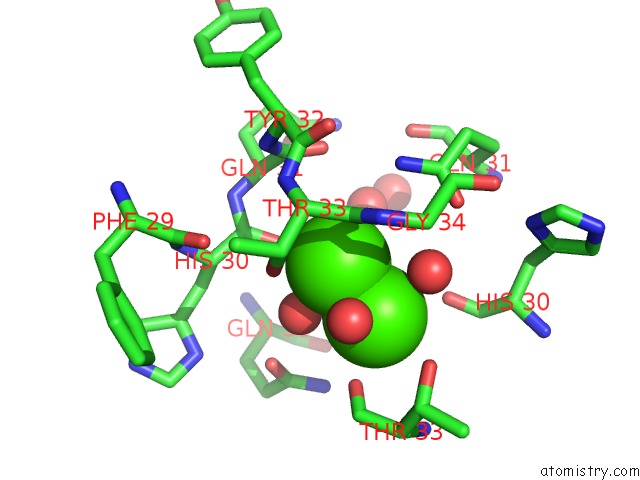
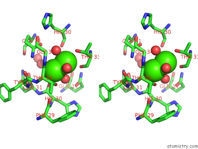
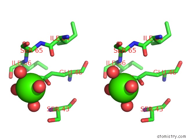
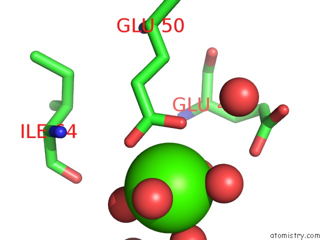
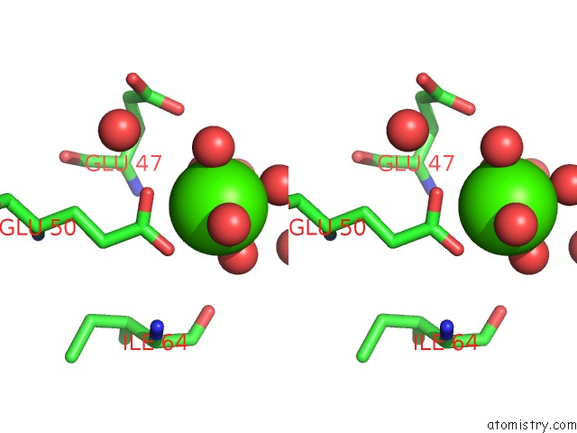
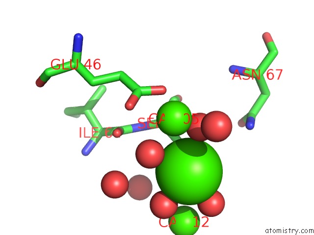
Calcium binding site 2 out of 13 in 2i52
Go back to
Calcium binding site 2 out
of 13 in the Crystal Structure of Protein PTO0218 From Picrophilus Torridus, Pfam DUF372
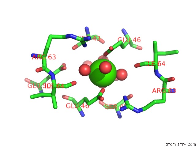
Mono view
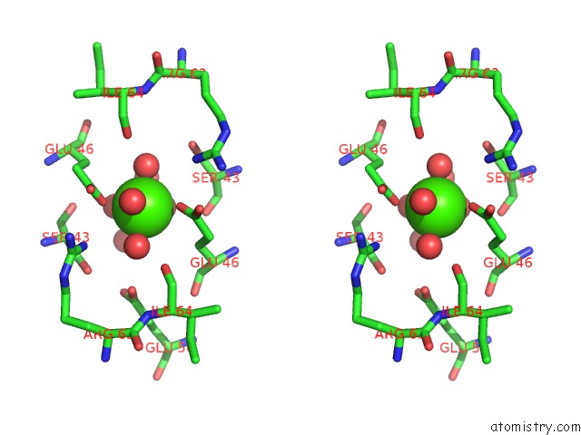
Stereo pair view
Mono view
Stereo pair view
A full contact list of Calcium with other atoms in the Ca binding
site number 2 of Crystal Structure of Protein PTO0218 From Picrophilus Torridus, Pfam DUF372 within 5.0Å range:
|
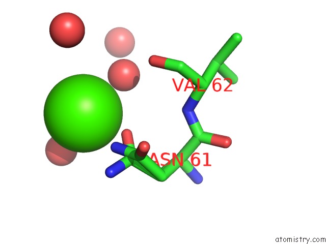
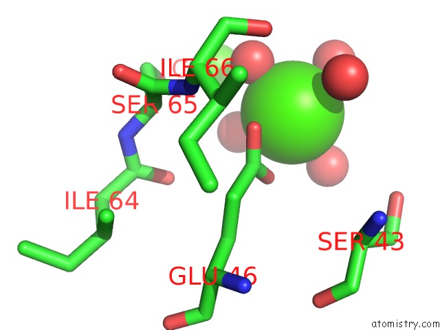
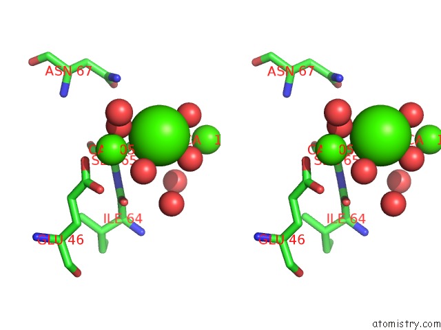
Calcium binding site 3 out of 13 in 2i52
Go back to
Calcium binding site 3 out
of 13 in the Crystal Structure of Protein PTO0218 From Picrophilus Torridus, Pfam DUF372
Mono view
Stereo pair view
Mono view
Stereo pair view
A full contact list of Calcium with other atoms in the Ca binding
site number 3 of Crystal Structure of Protein PTO0218 From Picrophilus Torridus, Pfam DUF372 within 5.0Å range:
|
Calcium binding site 4 out of 13 in 2i52
Go back to
Calcium binding site 4 out
of 13 in the Crystal Structure of Protein PTO0218 From Picrophilus Torridus, Pfam DUF372
Mono view
Stereo pair view
Mono view
Stereo pair view
A full contact list of Calcium with other atoms in the Ca binding
site number 4 of Crystal Structure of Protein PTO0218 From Picrophilus Torridus, Pfam DUF372 within 5.0Å range:
|
Calcium binding site 5 out of 13 in 2i52
Go back to
Calcium binding site 5 out
of 13 in the Crystal Structure of Protein PTO0218 From Picrophilus Torridus, Pfam DUF372
Mono view
Stereo pair view
Mono view
Stereo pair view
A full contact list of Calcium with other atoms in the Ca binding
site number 5 of Crystal Structure of Protein PTO0218 From Picrophilus Torridus, Pfam DUF372 within 5.0Å range:
|
Calcium binding site 6 out of 13 in 2i52
Go back to
Calcium binding site 6 out
of 13 in the Crystal Structure of Protein PTO0218 From Picrophilus Torridus, Pfam DUF372

Mono view
Stereo pair view
Mono view
Stereo pair view
A full contact list of Calcium with other atoms in the Ca binding
site number 6 of Crystal Structure of Protein PTO0218 From Picrophilus Torridus, Pfam DUF372 within 5.0Å range:
|
Calcium binding site 7 out of 13 in 2i52
Go back to
Calcium binding site 7 out
of 13 in the Crystal Structure of Protein PTO0218 From Picrophilus Torridus, Pfam DUF372
Mono view
Stereo pair view
Mono view
Stereo pair view
A full contact list of Calcium with other atoms in the Ca binding
site number 7 of Crystal Structure of Protein PTO0218 From Picrophilus Torridus, Pfam DUF372 within 5.0Å range:
|
Calcium binding site 8 out of 13 in 2i52
Go back to
Calcium binding site 8 out
of 13 in the Crystal Structure of Protein PTO0218 From Picrophilus Torridus, Pfam DUF372
Mono view
Stereo pair view
Mono view
Stereo pair view
A full contact list of Calcium with other atoms in the Ca binding
site number 8 of Crystal Structure of Protein PTO0218 From Picrophilus Torridus, Pfam DUF372 within 5.0Å range:
|
Calcium binding site 9 out of 13 in 2i52
Go back to
Calcium binding site 9 out
of 13 in the Crystal Structure of Protein PTO0218 From Picrophilus Torridus, Pfam DUF372
Mono view
Stereo pair view
Mono view
Stereo pair view
A full contact list of Calcium with other atoms in the Ca binding
site number 9 of Crystal Structure of Protein PTO0218 From Picrophilus Torridus, Pfam DUF372 within 5.0Å range:
|
Calcium binding site 10 out of 13 in 2i52
Go back to
Calcium binding site 10 out
of 13 in the Crystal Structure of Protein PTO0218 From Picrophilus Torridus, Pfam DUF372
Mono view
Stereo pair view
Mono view
Stereo pair view
A full contact list of Calcium with other atoms in the Ca binding
site number 10 of Crystal Structure of Protein PTO0218 From Picrophilus Torridus, Pfam DUF372 within 5.0Å range:
|
Reference:
U.A.Ramagopal,
J.Gilmore,
R.Toro,
K.T.Bain,
C.Mckenzie,
C.Reyes,
J.M.Sauder,
S.K.Burley,
S.C.Almo.
Structure of Hypothetical Protein PTO0218 From Picrophilus Torridus To Be Published.
Page generated: Tue Jul 8 06:08:45 2025
Last articles
Mn in 9LJUMn in 9LJW
Mn in 9LJS
Mn in 9LJR
Mn in 9LJT
Mn in 9LJV
Mg in 9UA2
Mg in 9R96
Mg in 9VM1
Mg in 9P01