Calcium »
PDB 2i5w-2imw »
2i5w »
Calcium in PDB 2i5w: Structure of HOGG1 Crosslinked to Dna Sampling A Normal G Adjacent to An Oxog
Enzymatic activity of Structure of HOGG1 Crosslinked to Dna Sampling A Normal G Adjacent to An Oxog
All present enzymatic activity of Structure of HOGG1 Crosslinked to Dna Sampling A Normal G Adjacent to An Oxog:
4.2.99.18;
4.2.99.18;
Protein crystallography data
The structure of Structure of HOGG1 Crosslinked to Dna Sampling A Normal G Adjacent to An Oxog, PDB code: 2i5w
was solved by
A.Banerjee,
G.L.Verdine,
with X-Ray Crystallography technique. A brief refinement statistics is given in the table below:
| Resolution Low / High (Å) | 45.76 / 2.60 |
| Space group | P 65 2 2 |
| Cell size a, b, c (Å), α, β, γ (°) | 91.524, 91.524, 211.848, 90.00, 90.00, 120.00 |
| R / Rfree (%) | 22.6 / 26.6 |
Calcium Binding Sites:
The binding sites of Calcium atom in the Structure of HOGG1 Crosslinked to Dna Sampling A Normal G Adjacent to An Oxog
(pdb code 2i5w). This binding sites where shown within
5.0 Angstroms radius around Calcium atom.
In total 2 binding sites of Calcium where determined in the Structure of HOGG1 Crosslinked to Dna Sampling A Normal G Adjacent to An Oxog, PDB code: 2i5w:
Jump to Calcium binding site number: 1; 2;
In total 2 binding sites of Calcium where determined in the Structure of HOGG1 Crosslinked to Dna Sampling A Normal G Adjacent to An Oxog, PDB code: 2i5w:
Jump to Calcium binding site number: 1; 2;
Calcium binding site 1 out of 2 in 2i5w
Go back to
Calcium binding site 1 out
of 2 in the Structure of HOGG1 Crosslinked to Dna Sampling A Normal G Adjacent to An Oxog
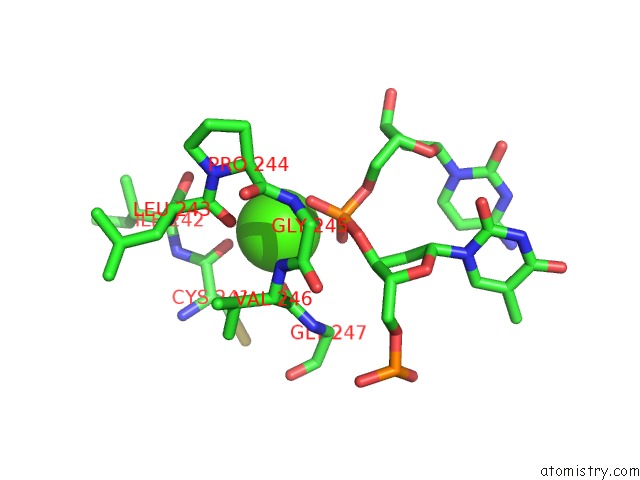
Mono view
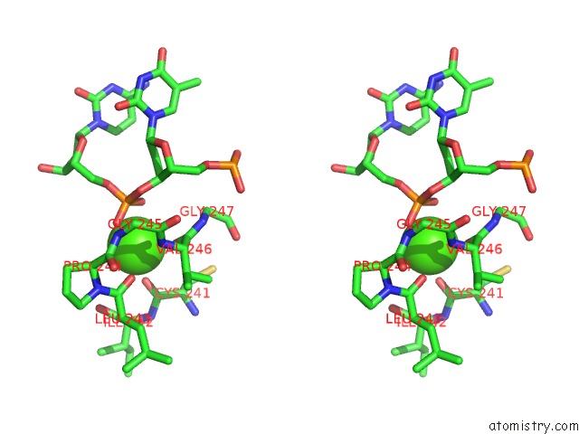
Stereo pair view
Mono view
Stereo pair view
A full contact list of Calcium with other atoms in the Ca binding
site number 1 of Structure of HOGG1 Crosslinked to Dna Sampling A Normal G Adjacent to An Oxog within 5.0Å range:
|
Calcium binding site 2 out of 2 in 2i5w
Go back to
Calcium binding site 2 out
of 2 in the Structure of HOGG1 Crosslinked to Dna Sampling A Normal G Adjacent to An Oxog
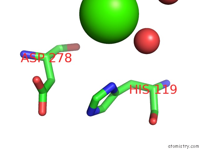
Mono view
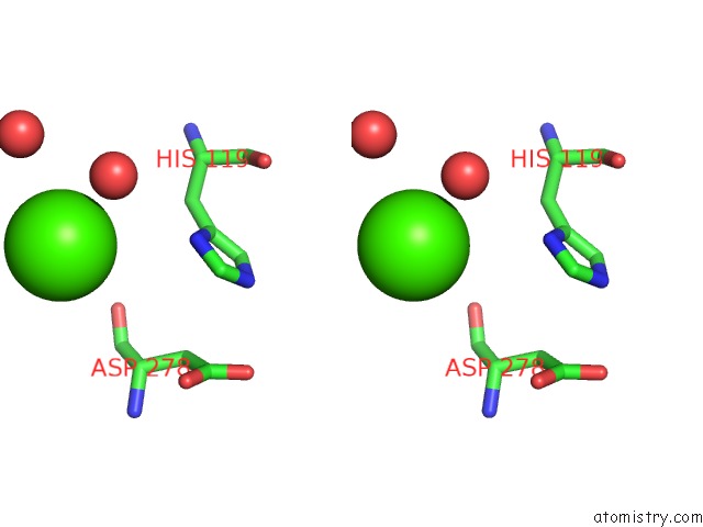
Stereo pair view
Mono view
Stereo pair view
A full contact list of Calcium with other atoms in the Ca binding
site number 2 of Structure of HOGG1 Crosslinked to Dna Sampling A Normal G Adjacent to An Oxog within 5.0Å range:
|
Reference:
A.Banerjee,
G.L.Verdine.
A Nucleobase Lesion Remodels the Interaction of Its Normal Neighbor in A Dna Glycosylase Complex. Proc.Natl.Acad.Sci.Usa V. 103 15020 2006.
ISSN: ISSN 0027-8424
PubMed: 17015827
DOI: 10.1073/PNAS.0603644103
Page generated: Tue Jul 8 06:10:33 2025
ISSN: ISSN 0027-8424
PubMed: 17015827
DOI: 10.1073/PNAS.0603644103
Last articles
K in 6R2TK in 6R2R
K in 6R2P
K in 6R2I
K in 6QML
K in 6QXA
K in 6R0K
K in 6R15
K in 6QJO
K in 6QD4