Calcium »
PDB 2i5w-2imw »
2id4 »
Calcium in PDB 2id4: The 1.9 A Structure of KEX2 in Complex with An Ac-R-E-R-K-Chloromethyl Ketone Inhibitor.
Enzymatic activity of The 1.9 A Structure of KEX2 in Complex with An Ac-R-E-R-K-Chloromethyl Ketone Inhibitor.
All present enzymatic activity of The 1.9 A Structure of KEX2 in Complex with An Ac-R-E-R-K-Chloromethyl Ketone Inhibitor.:
3.4.21.61;
3.4.21.61;
Protein crystallography data
The structure of The 1.9 A Structure of KEX2 in Complex with An Ac-R-E-R-K-Chloromethyl Ketone Inhibitor., PDB code: 2id4
was solved by
J.L.Wheatley,
T.Holyoak,
with X-Ray Crystallography technique. A brief refinement statistics is given in the table below:
| Resolution Low / High (Å) | 40.79 / 1.90 |
| Space group | P 65 2 2 |
| Cell size a, b, c (Å), α, β, γ (°) | 112.851, 112.851, 370.165, 90.00, 90.00, 120.00 |
| R / Rfree (%) | 17.7 / 20.6 |
Other elements in 2id4:
The structure of The 1.9 A Structure of KEX2 in Complex with An Ac-R-E-R-K-Chloromethyl Ketone Inhibitor. also contains other interesting chemical elements:
| Sodium | (Na) | 2 atoms |
Calcium Binding Sites:
The binding sites of Calcium atom in the The 1.9 A Structure of KEX2 in Complex with An Ac-R-E-R-K-Chloromethyl Ketone Inhibitor.
(pdb code 2id4). This binding sites where shown within
5.0 Angstroms radius around Calcium atom.
In total 4 binding sites of Calcium where determined in the The 1.9 A Structure of KEX2 in Complex with An Ac-R-E-R-K-Chloromethyl Ketone Inhibitor., PDB code: 2id4:
Jump to Calcium binding site number: 1; 2; 3; 4;
In total 4 binding sites of Calcium where determined in the The 1.9 A Structure of KEX2 in Complex with An Ac-R-E-R-K-Chloromethyl Ketone Inhibitor., PDB code: 2id4:
Jump to Calcium binding site number: 1; 2; 3; 4;
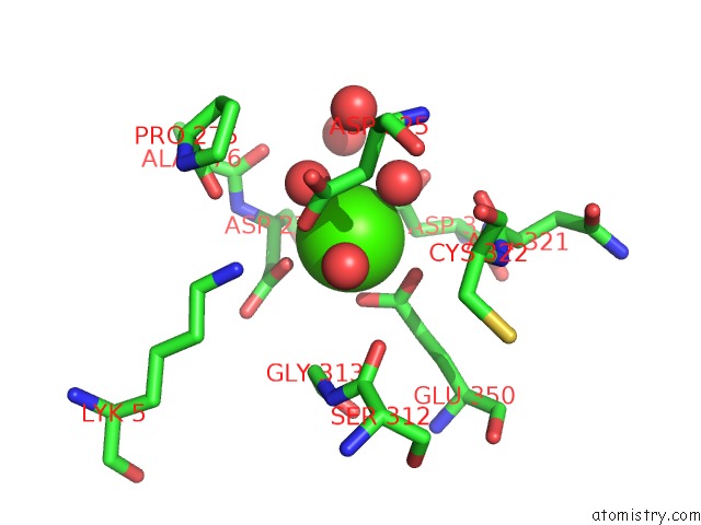
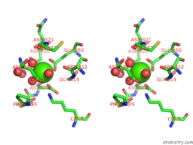
Calcium binding site 1 out of 4 in 2id4
Go back to
Calcium binding site 1 out
of 4 in the The 1.9 A Structure of KEX2 in Complex with An Ac-R-E-R-K-Chloromethyl Ketone Inhibitor.
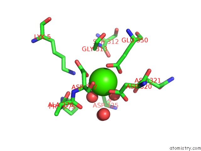
Mono view
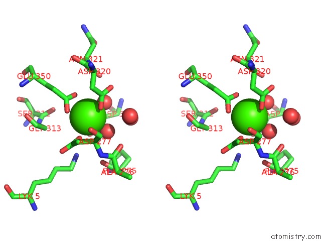
Stereo pair view
Mono view
Stereo pair view
A full contact list of Calcium with other atoms in the Ca binding
site number 1 of The 1.9 A Structure of KEX2 in Complex with An Ac-R-E-R-K-Chloromethyl Ketone Inhibitor. within 5.0Å range:
|
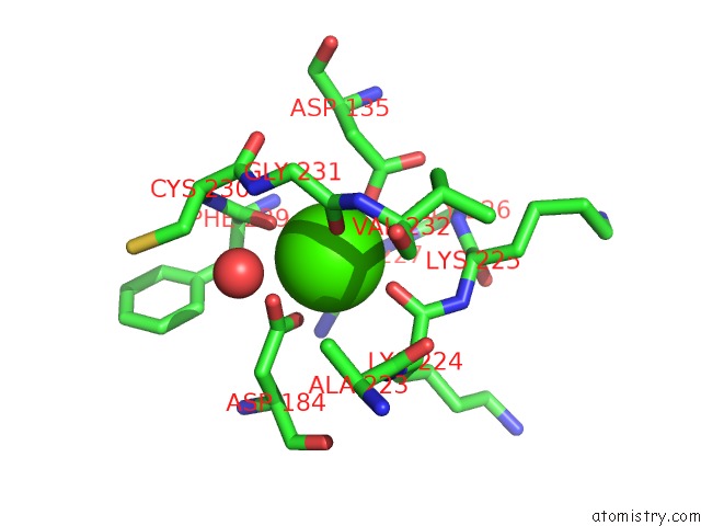
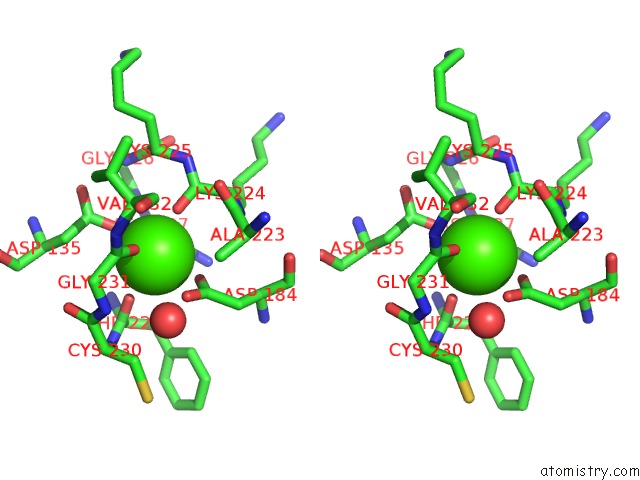
Calcium binding site 2 out of 4 in 2id4
Go back to
Calcium binding site 2 out
of 4 in the The 1.9 A Structure of KEX2 in Complex with An Ac-R-E-R-K-Chloromethyl Ketone Inhibitor.
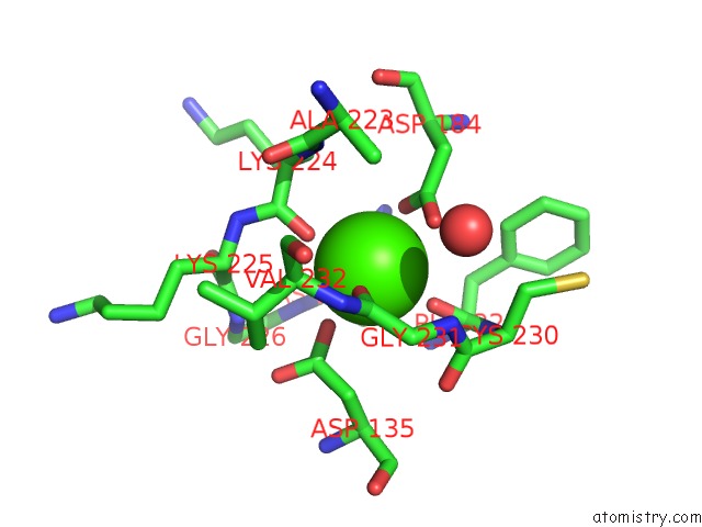
Mono view
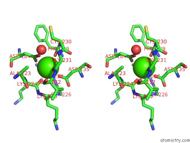
Stereo pair view
Mono view
Stereo pair view
A full contact list of Calcium with other atoms in the Ca binding
site number 2 of The 1.9 A Structure of KEX2 in Complex with An Ac-R-E-R-K-Chloromethyl Ketone Inhibitor. within 5.0Å range:
|
Calcium binding site 3 out of 4 in 2id4
Go back to
Calcium binding site 3 out
of 4 in the The 1.9 A Structure of KEX2 in Complex with An Ac-R-E-R-K-Chloromethyl Ketone Inhibitor.
Mono view
Stereo pair view
Mono view
Stereo pair view
A full contact list of Calcium with other atoms in the Ca binding
site number 3 of The 1.9 A Structure of KEX2 in Complex with An Ac-R-E-R-K-Chloromethyl Ketone Inhibitor. within 5.0Å range:
|
Calcium binding site 4 out of 4 in 2id4
Go back to
Calcium binding site 4 out
of 4 in the The 1.9 A Structure of KEX2 in Complex with An Ac-R-E-R-K-Chloromethyl Ketone Inhibitor.
Mono view
Stereo pair view
Mono view
Stereo pair view
A full contact list of Calcium with other atoms in the Ca binding
site number 4 of The 1.9 A Structure of KEX2 in Complex with An Ac-R-E-R-K-Chloromethyl Ketone Inhibitor. within 5.0Å range:
|
Reference:
J.L.Wheatley,
T.Holyoak.
Differential P1 Arginine and Lysine Recognition in the Prototypical Proprotein Convertase KEX2. Proc.Natl.Acad.Sci.Usa V. 104 6626 2007.
ISSN: ISSN 0027-8424
PubMed: 17426142
DOI: 10.1073/PNAS.0701983104
Page generated: Tue Jul 8 06:13:08 2025
ISSN: ISSN 0027-8424
PubMed: 17426142
DOI: 10.1073/PNAS.0701983104
Last articles
K in 8RZ6K in 8RMH
K in 8RS5
K in 8R6F
K in 8RSN
K in 8RPK
K in 8ROO
K in 8RIU
K in 8RGR
K in 8RGT