Calcium »
PDB 2i5w-2imw »
2idj »
Calcium in PDB 2idj: Crystal Structure of Rat Glycine N-Methyltransferase Apoprotein, Monoclinic Form
Enzymatic activity of Crystal Structure of Rat Glycine N-Methyltransferase Apoprotein, Monoclinic Form
All present enzymatic activity of Crystal Structure of Rat Glycine N-Methyltransferase Apoprotein, Monoclinic Form:
2.1.1.20;
2.1.1.20;
Protein crystallography data
The structure of Crystal Structure of Rat Glycine N-Methyltransferase Apoprotein, Monoclinic Form, PDB code: 2idj
was solved by
Z.Luka,
S.Pakhomova,
L.V.Loukachevitch,
M.Egli,
M.E.Newcomer,
C.Wagner,
with X-Ray Crystallography technique. A brief refinement statistics is given in the table below:
| Resolution Low / High (Å) | 39.11 / 2.35 |
| Space group | P 1 21 1 |
| Cell size a, b, c (Å), α, β, γ (°) | 57.869, 85.223, 131.861, 90.00, 91.40, 90.00 |
| R / Rfree (%) | 22.2 / 27.2 |
Calcium Binding Sites:
The binding sites of Calcium atom in the Crystal Structure of Rat Glycine N-Methyltransferase Apoprotein, Monoclinic Form
(pdb code 2idj). This binding sites where shown within
5.0 Angstroms radius around Calcium atom.
In total 2 binding sites of Calcium where determined in the Crystal Structure of Rat Glycine N-Methyltransferase Apoprotein, Monoclinic Form, PDB code: 2idj:
Jump to Calcium binding site number: 1; 2;
In total 2 binding sites of Calcium where determined in the Crystal Structure of Rat Glycine N-Methyltransferase Apoprotein, Monoclinic Form, PDB code: 2idj:
Jump to Calcium binding site number: 1; 2;
Calcium binding site 1 out of 2 in 2idj
Go back to
Calcium binding site 1 out
of 2 in the Crystal Structure of Rat Glycine N-Methyltransferase Apoprotein, Monoclinic Form
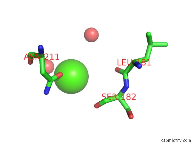
Mono view
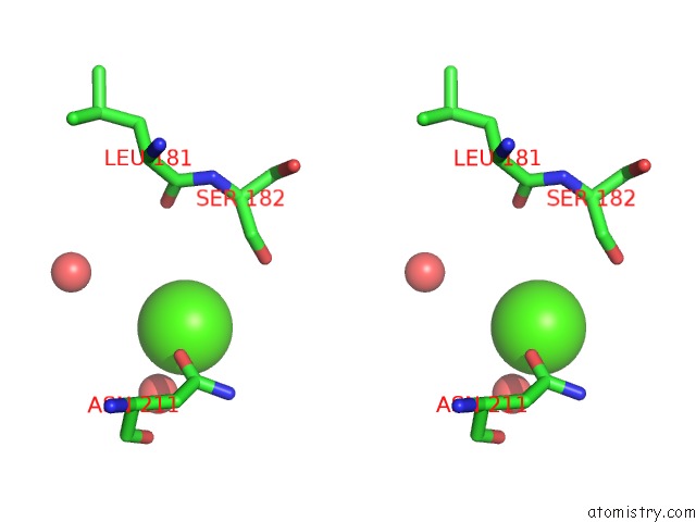
Stereo pair view
Mono view
Stereo pair view
A full contact list of Calcium with other atoms in the Ca binding
site number 1 of Crystal Structure of Rat Glycine N-Methyltransferase Apoprotein, Monoclinic Form within 5.0Å range:
|
Calcium binding site 2 out of 2 in 2idj
Go back to
Calcium binding site 2 out
of 2 in the Crystal Structure of Rat Glycine N-Methyltransferase Apoprotein, Monoclinic Form
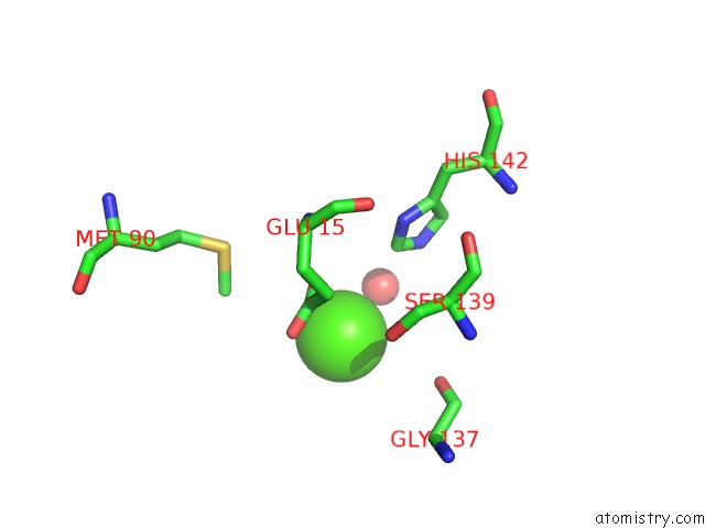
Mono view
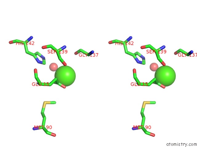
Stereo pair view
Mono view
Stereo pair view
A full contact list of Calcium with other atoms in the Ca binding
site number 2 of Crystal Structure of Rat Glycine N-Methyltransferase Apoprotein, Monoclinic Form within 5.0Å range:
|
Reference:
Z.Luka,
S.Pakhomova,
L.V.Loukachevitch,
M.Egli,
M.E.Newcomer,
C.Wagner.
5-Methyltetrahydrofolate Is Bound in Intersubunit Areas of Rat Liver Folate-Binding Protein Glycine N-Methyltransferase. J.Biol.Chem. V. 282 4069 2007.
ISSN: ISSN 0021-9258
PubMed: 17158459
DOI: 10.1074/JBC.M610384200
Page generated: Tue Jul 8 06:13:16 2025
ISSN: ISSN 0021-9258
PubMed: 17158459
DOI: 10.1074/JBC.M610384200
Last articles
K in 5E33K in 5E2Q
K in 5E1A
K in 5DWX
K in 5DUN
K in 5DSX
K in 5DRY
K in 5DRT
K in 5DMJ
K in 5DQK