Calcium »
PDB 2j3u-2jdh »
2jam »
Calcium in PDB 2jam: Crystal Structure of Human Calmodulin-Dependent Protein Kinase I G
Enzymatic activity of Crystal Structure of Human Calmodulin-Dependent Protein Kinase I G
All present enzymatic activity of Crystal Structure of Human Calmodulin-Dependent Protein Kinase I G:
2.7.11.17;
2.7.11.17;
Protein crystallography data
The structure of Crystal Structure of Human Calmodulin-Dependent Protein Kinase I G, PDB code: 2jam
was solved by
J.E.Debreczeni,
A.Bullock,
T.Keates,
F.H.Niesen,
E.Salah,
L.Shrestha,
C.Smee,
F.Sobott,
A.C.W.Pike,
G.Bunkoczi,
F.Von Delft,
A.Turnbull,
J.Weigelt,
C.H.Arrowsmith,
A.Edwards,
M.Sundstrom,
S.Knapp,
with X-Ray Crystallography technique. A brief refinement statistics is given in the table below:
| Resolution Low / High (Å) | 56.89 / 1.70 |
| Space group | P 1 |
| Cell size a, b, c (Å), α, β, γ (°) | 51.666, 53.736, 64.177, 65.45, 71.91, 73.84 |
| R / Rfree (%) | 15.5 / 23.1 |
Other elements in 2jam:
The structure of Crystal Structure of Human Calmodulin-Dependent Protein Kinase I G also contains other interesting chemical elements:
| Chlorine | (Cl) | 2 atoms |
Calcium Binding Sites:
The binding sites of Calcium atom in the Crystal Structure of Human Calmodulin-Dependent Protein Kinase I G
(pdb code 2jam). This binding sites where shown within
5.0 Angstroms radius around Calcium atom.
In total only one binding site of Calcium was determined in the Crystal Structure of Human Calmodulin-Dependent Protein Kinase I G, PDB code: 2jam:
In total only one binding site of Calcium was determined in the Crystal Structure of Human Calmodulin-Dependent Protein Kinase I G, PDB code: 2jam:
Calcium binding site 1 out of 1 in 2jam
Go back to
Calcium binding site 1 out
of 1 in the Crystal Structure of Human Calmodulin-Dependent Protein Kinase I G
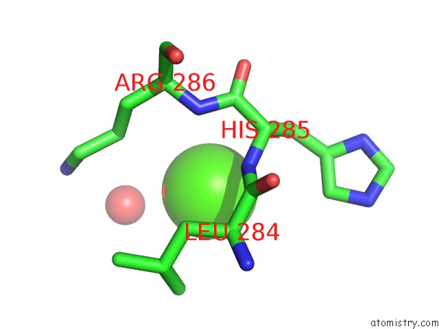
Mono view
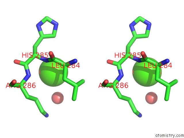
Stereo pair view
Mono view
Stereo pair view
A full contact list of Calcium with other atoms in the Ca binding
site number 1 of Crystal Structure of Human Calmodulin-Dependent Protein Kinase I G within 5.0Å range:
|
Reference:
J.E.Debreczeni,
A.Bullock,
T.Keates,
F.H.Niesen,
E.Salah,
L.Shrestha,
C.Smee,
F.Sobott,
A.C.W.Pike,
G.Bunkoczi,
F.Von Delft,
A.Turnbull,
J.Weigelt,
C.H.Arrowsmith,
A.Edwards,
M.Sundstrom,
S.Knapp.
Crystal Structure of Human Calmodulin-Dependent Protein Kinase I G To Be Published.
Page generated: Tue Jul 8 06:33:46 2025
Last articles
Na in 2QB4Na in 2QD6
Na in 2QCI
Na in 2QA0
Na in 2Q95
Na in 2Q96
Na in 2Q94
Na in 2Q93
Na in 2Q8X
Na in 2Q72