Calcium »
PDB 2m0k-2n8y »
2mcm »
Calcium in PDB 2mcm: Macromomycin
Protein crystallography data
The structure of Macromomycin, PDB code: 2mcm
was solved by
P.Van Roey,
with X-Ray Crystallography technique. A brief refinement statistics is given in the table below:
| Resolution Low / High (Å) | 10.00 / 1.50 |
| Space group | P 1 21 1 |
| Cell size a, b, c (Å), α, β, γ (°) | 36.290, 35.580, 38.040, 90.00, 99.59, 90.00 |
| R / Rfree (%) | n/a / n/a |
Calcium Binding Sites:
The binding sites of Calcium atom in the Macromomycin
(pdb code 2mcm). This binding sites where shown within
5.0 Angstroms radius around Calcium atom.
In total only one binding site of Calcium was determined in the Macromomycin, PDB code: 2mcm:
In total only one binding site of Calcium was determined in the Macromomycin, PDB code: 2mcm:
Calcium binding site 1 out of 1 in 2mcm
Go back to
Calcium binding site 1 out
of 1 in the Macromomycin
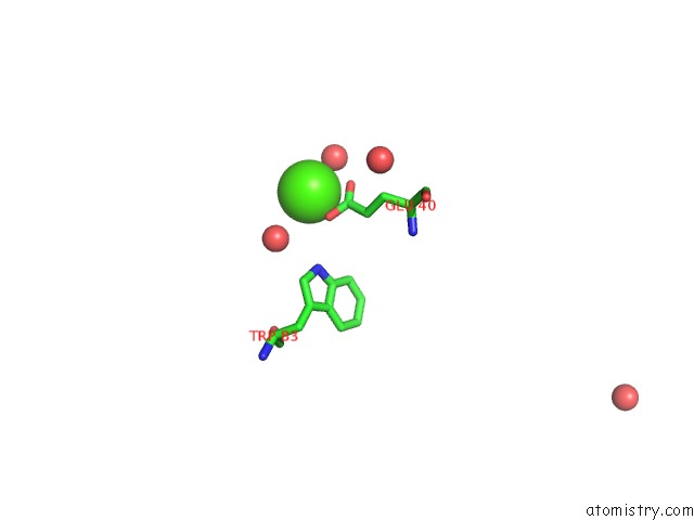
Mono view
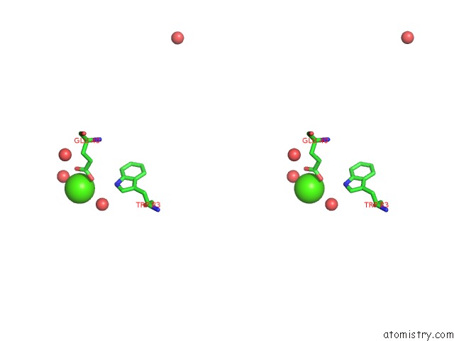
Stereo pair view
Mono view
Stereo pair view
A full contact list of Calcium with other atoms in the Ca binding
site number 1 of Macromomycin within 5.0Å range:
|
Reference:
P.Van Roey,
T.A.Beerman.
Crystal Structure Analysis of Auromomycin Apoprotein (Macromomycin) Shows Importance of Protein Side Chains to Chromophore Binding Selectivity. Proc.Natl.Acad.Sci.Usa V. 86 6587 1989.
ISSN: ISSN 0027-8424
PubMed: 2771945
DOI: 10.1073/PNAS.86.17.6587
Page generated: Tue Jul 8 07:04:51 2025
ISSN: ISSN 0027-8424
PubMed: 2771945
DOI: 10.1073/PNAS.86.17.6587
Last articles
Mg in 8CGIMg in 8CF8
Mg in 8CGA
Mg in 8CEP
Mg in 8CF1
Mg in 8CAH
Mg in 8CE5
Mg in 8CDQ
Mg in 8CE2
Mg in 8CCO