Calcium »
PDB 2nce-2o86 »
2nof »
Calcium in PDB 2nof: Structure of Q315F Human 8-Oxoguanine Glycosylase Proximal Crosslink to 8-Oxoguanine Dna
Enzymatic activity of Structure of Q315F Human 8-Oxoguanine Glycosylase Proximal Crosslink to 8-Oxoguanine Dna
All present enzymatic activity of Structure of Q315F Human 8-Oxoguanine Glycosylase Proximal Crosslink to 8-Oxoguanine Dna:
4.2.99.18;
4.2.99.18;
Protein crystallography data
The structure of Structure of Q315F Human 8-Oxoguanine Glycosylase Proximal Crosslink to 8-Oxoguanine Dna, PDB code: 2nof
was solved by
C.T.Radom,
A.Banerjee,
G.L.Verdine,
with X-Ray Crystallography technique. A brief refinement statistics is given in the table below:
| Resolution Low / High (Å) | 50.00 / 2.35 |
| Space group | P 65 2 2 |
| Cell size a, b, c (Å), α, β, γ (°) | 92.267, 92.267, 211.128, 90.00, 90.00, 120.00 |
| R / Rfree (%) | 22.7 / 27 |
Calcium Binding Sites:
The binding sites of Calcium atom in the Structure of Q315F Human 8-Oxoguanine Glycosylase Proximal Crosslink to 8-Oxoguanine Dna
(pdb code 2nof). This binding sites where shown within
5.0 Angstroms radius around Calcium atom.
In total only one binding site of Calcium was determined in the Structure of Q315F Human 8-Oxoguanine Glycosylase Proximal Crosslink to 8-Oxoguanine Dna, PDB code: 2nof:
In total only one binding site of Calcium was determined in the Structure of Q315F Human 8-Oxoguanine Glycosylase Proximal Crosslink to 8-Oxoguanine Dna, PDB code: 2nof:
Calcium binding site 1 out of 1 in 2nof
Go back to
Calcium binding site 1 out
of 1 in the Structure of Q315F Human 8-Oxoguanine Glycosylase Proximal Crosslink to 8-Oxoguanine Dna
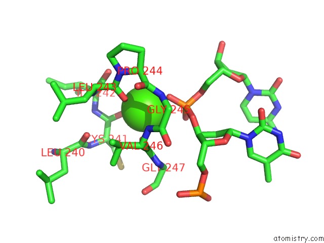
Mono view
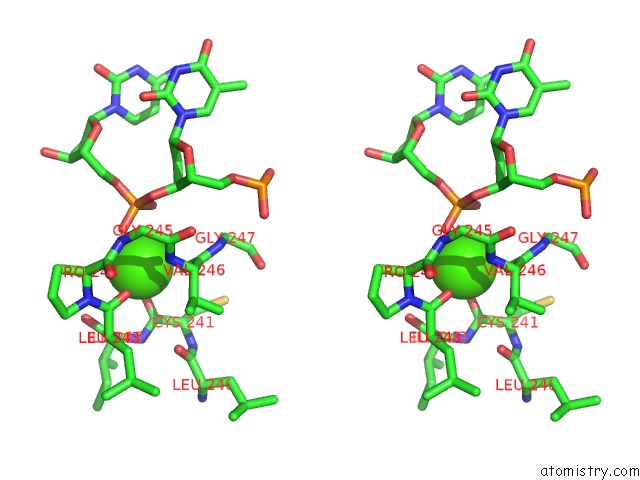
Stereo pair view
Mono view
Stereo pair view
A full contact list of Calcium with other atoms in the Ca binding
site number 1 of Structure of Q315F Human 8-Oxoguanine Glycosylase Proximal Crosslink to 8-Oxoguanine Dna within 5.0Å range:
|
Reference:
C.T.Radom,
A.Banerjee,
G.L.Verdine.
Structural Characterization of Human 8-Oxoguanine Dna Glycosylase Variants Bearing Active Site Mutations. J.Biol.Chem. V. 282 9182 2007.
ISSN: ISSN 0021-9258
PubMed: 17114185
DOI: 10.1074/JBC.M608989200
Page generated: Tue Jul 8 07:11:41 2025
ISSN: ISSN 0021-9258
PubMed: 17114185
DOI: 10.1074/JBC.M608989200
Last articles
Mg in 5LTKMg in 5LTF
Mg in 5LSE
Mg in 5LTJ
Mg in 5LTA
Mg in 5LS9
Mg in 5LSA
Mg in 5LS0
Mg in 5LS7
Mg in 5LRT