Calcium »
PDB 2nce-2o86 »
2np0 »
Calcium in PDB 2np0: Crystal Structure of the Botulinum Neurotoxin Type B Complexed with Synaptotagamin-II Ectodomain
Enzymatic activity of Crystal Structure of the Botulinum Neurotoxin Type B Complexed with Synaptotagamin-II Ectodomain
All present enzymatic activity of Crystal Structure of the Botulinum Neurotoxin Type B Complexed with Synaptotagamin-II Ectodomain:
3.4.24.69;
3.4.24.69;
Protein crystallography data
The structure of Crystal Structure of the Botulinum Neurotoxin Type B Complexed with Synaptotagamin-II Ectodomain, PDB code: 2np0
was solved by
Q.Chai,
J.W.Arndt,
R.C.Stevens,
with X-Ray Crystallography technique. A brief refinement statistics is given in the table below:
| Resolution Low / High (Å) | 40.00 / 2.62 |
| Space group | C 2 2 21 |
| Cell size a, b, c (Å), α, β, γ (°) | 170.415, 351.971, 101.968, 90.00, 90.00, 90.00 |
| R / Rfree (%) | 18.6 / 22.7 |
Other elements in 2np0:
The structure of Crystal Structure of the Botulinum Neurotoxin Type B Complexed with Synaptotagamin-II Ectodomain also contains other interesting chemical elements:
| Chlorine | (Cl) | 1 atom |
| Zinc | (Zn) | 1 atom |
Calcium Binding Sites:
The binding sites of Calcium atom in the Crystal Structure of the Botulinum Neurotoxin Type B Complexed with Synaptotagamin-II Ectodomain
(pdb code 2np0). This binding sites where shown within
5.0 Angstroms radius around Calcium atom.
In total 3 binding sites of Calcium where determined in the Crystal Structure of the Botulinum Neurotoxin Type B Complexed with Synaptotagamin-II Ectodomain, PDB code: 2np0:
Jump to Calcium binding site number: 1; 2; 3;
In total 3 binding sites of Calcium where determined in the Crystal Structure of the Botulinum Neurotoxin Type B Complexed with Synaptotagamin-II Ectodomain, PDB code: 2np0:
Jump to Calcium binding site number: 1; 2; 3;
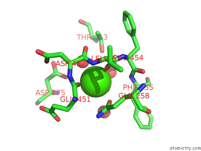
Calcium binding site 1 out of 3 in 2np0
Go back to
Calcium binding site 1 out
of 3 in the Crystal Structure of the Botulinum Neurotoxin Type B Complexed with Synaptotagamin-II Ectodomain
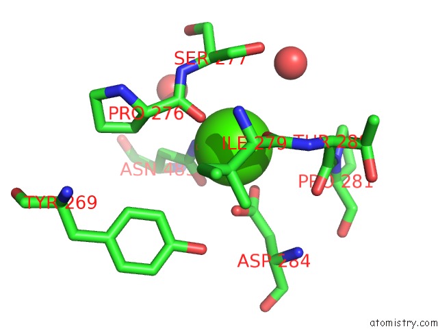
Mono view
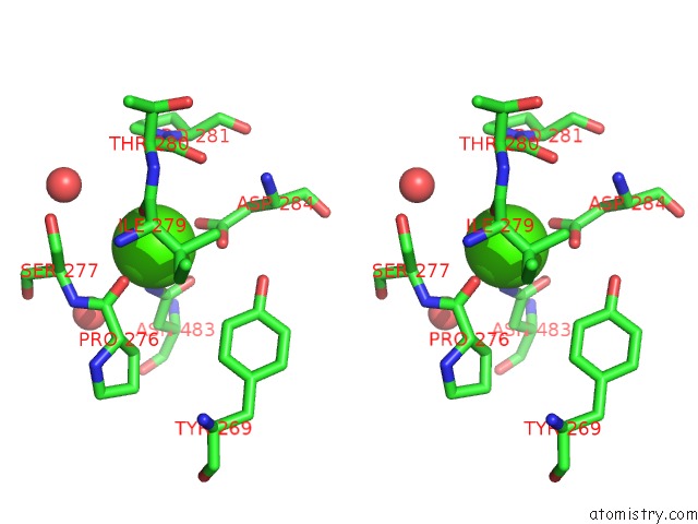
Stereo pair view
Mono view
Stereo pair view
A full contact list of Calcium with other atoms in the Ca binding
site number 1 of Crystal Structure of the Botulinum Neurotoxin Type B Complexed with Synaptotagamin-II Ectodomain within 5.0Å range:
|
Calcium binding site 2 out of 3 in 2np0
Go back to
Calcium binding site 2 out
of 3 in the Crystal Structure of the Botulinum Neurotoxin Type B Complexed with Synaptotagamin-II Ectodomain
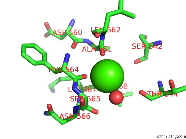
Mono view
Stereo pair view
Mono view
Stereo pair view
A full contact list of Calcium with other atoms in the Ca binding
site number 2 of Crystal Structure of the Botulinum Neurotoxin Type B Complexed with Synaptotagamin-II Ectodomain within 5.0Å range:
|
Calcium binding site 3 out of 3 in 2np0
Go back to
Calcium binding site 3 out
of 3 in the Crystal Structure of the Botulinum Neurotoxin Type B Complexed with Synaptotagamin-II Ectodomain
Mono view
Stereo pair view
Mono view
Stereo pair view
A full contact list of Calcium with other atoms in the Ca binding
site number 3 of Crystal Structure of the Botulinum Neurotoxin Type B Complexed with Synaptotagamin-II Ectodomain within 5.0Å range:
|
Reference:
Q.Chai,
J.W.Arndt,
M.Dong,
W.H.Tepp,
E.A.Johnson,
E.R.Chapman,
R.C.Stevens.
Structural Basis of Cell Surface Receptor Recognition By Botulinum Neurotoxin B. Nature V. 444 1096 2006.
ISSN: ISSN 0028-0836
PubMed: 17167418
DOI: 10.1038/NATURE05411
Page generated: Tue Jul 8 07:12:19 2025
ISSN: ISSN 0028-0836
PubMed: 17167418
DOI: 10.1038/NATURE05411
Last articles
K in 1TYYK in 1TI7
K in 1THZ
K in 1T9D
K in 1TTQ
K in 1TGY
K in 1TA9
K in 1TGV
K in 1T9C
K in 1T60