Calcium »
PDB 2o8o-2ovu »
2oa8 »
Calcium in PDB 2oa8: Crystal Structure of MTREX1 with Ssdna
Enzymatic activity of Crystal Structure of MTREX1 with Ssdna
All present enzymatic activity of Crystal Structure of MTREX1 with Ssdna:
3.1.11.2;
3.1.11.2;
Protein crystallography data
The structure of Crystal Structure of MTREX1 with Ssdna, PDB code: 2oa8
was solved by
U.De Silva,
T.Hollis,
with X-Ray Crystallography technique. A brief refinement statistics is given in the table below:
| Resolution Low / High (Å) | 61.90 / 2.10 |
| Space group | P 1 21 1 |
| Cell size a, b, c (Å), α, β, γ (°) | 64.850, 57.141, 68.470, 90.00, 107.47, 90.00 |
| R / Rfree (%) | 19.2 / 25.9 |
Calcium Binding Sites:
The binding sites of Calcium atom in the Crystal Structure of MTREX1 with Ssdna
(pdb code 2oa8). This binding sites where shown within
5.0 Angstroms radius around Calcium atom.
In total 4 binding sites of Calcium where determined in the Crystal Structure of MTREX1 with Ssdna, PDB code: 2oa8:
Jump to Calcium binding site number: 1; 2; 3; 4;
In total 4 binding sites of Calcium where determined in the Crystal Structure of MTREX1 with Ssdna, PDB code: 2oa8:
Jump to Calcium binding site number: 1; 2; 3; 4;
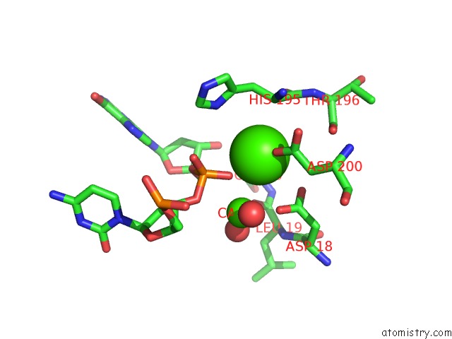
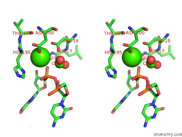
Calcium binding site 1 out of 4 in 2oa8
Go back to
Calcium binding site 1 out
of 4 in the Crystal Structure of MTREX1 with Ssdna
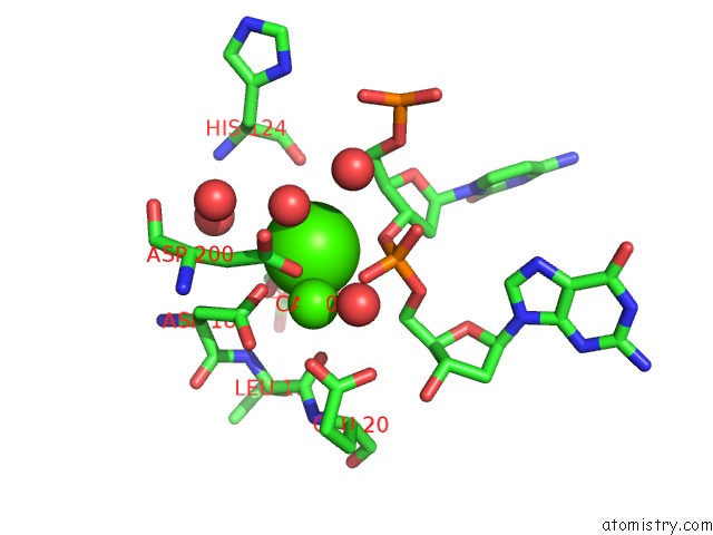
Mono view
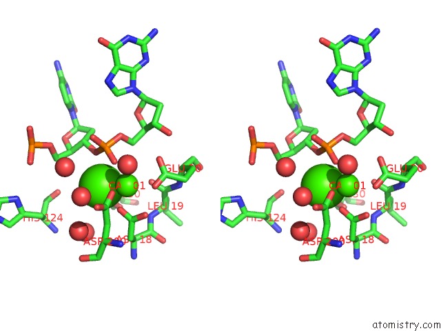
Stereo pair view
Mono view
Stereo pair view
A full contact list of Calcium with other atoms in the Ca binding
site number 1 of Crystal Structure of MTREX1 with Ssdna within 5.0Å range:
|
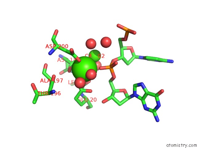
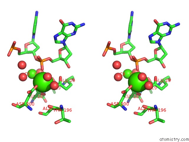
Calcium binding site 2 out of 4 in 2oa8
Go back to
Calcium binding site 2 out
of 4 in the Crystal Structure of MTREX1 with Ssdna
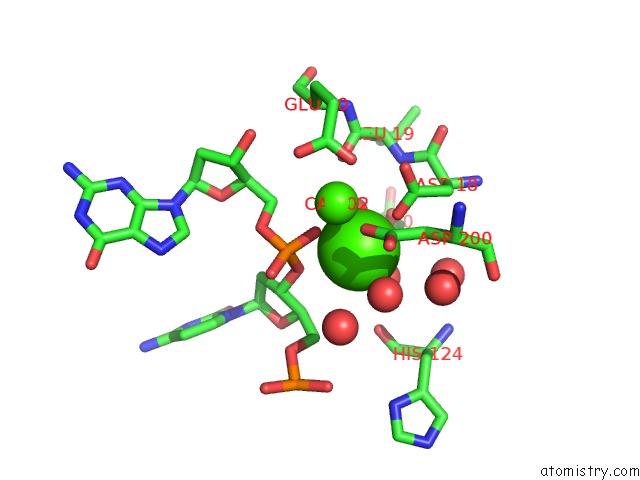
Mono view
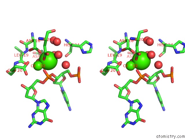
Stereo pair view
Mono view
Stereo pair view
A full contact list of Calcium with other atoms in the Ca binding
site number 2 of Crystal Structure of MTREX1 with Ssdna within 5.0Å range:
|
Calcium binding site 3 out of 4 in 2oa8
Go back to
Calcium binding site 3 out
of 4 in the Crystal Structure of MTREX1 with Ssdna
Mono view
Stereo pair view
Mono view
Stereo pair view
A full contact list of Calcium with other atoms in the Ca binding
site number 3 of Crystal Structure of MTREX1 with Ssdna within 5.0Å range:
|
Calcium binding site 4 out of 4 in 2oa8
Go back to
Calcium binding site 4 out
of 4 in the Crystal Structure of MTREX1 with Ssdna
Mono view
Stereo pair view
Mono view
Stereo pair view
A full contact list of Calcium with other atoms in the Ca binding
site number 4 of Crystal Structure of MTREX1 with Ssdna within 5.0Å range:
|
Reference:
U.De Silva,
S.Choudhury,
S.L.Bailey,
S.Harvey,
F.W.Perrino,
T.Hollis.
The Crystal Structure of TREX1 Explains the 3' Nucleotide Specificity and Reveals A Polyproline II Helix For Protein Partnering. J.Biol.Chem. V. 282 10537 2007.
ISSN: ISSN 0021-9258
PubMed: 17293595
DOI: 10.1074/JBC.M700039200
Page generated: Tue Jul 8 07:18:18 2025
ISSN: ISSN 0021-9258
PubMed: 17293595
DOI: 10.1074/JBC.M700039200
Last articles
Mg in 6C8LMg in 6C8J
Mg in 6C8K
Mg in 6C8I
Mg in 6C7J
Mg in 6C7Y
Mg in 6C7I
Mg in 6C4H
Mg in 6C7G
Mg in 6C7F