Calcium »
PDB 2o8o-2ovu »
2oan »
Calcium in PDB 2oan: Structure of Oxidized Beta-Actin
Protein crystallography data
The structure of Structure of Oxidized Beta-Actin, PDB code: 2oan
was solved by
F.Schmitzberger,
I.Lassing,
P.Nordlund,
U.Lindberg,
with X-Ray Crystallography technique. A brief refinement statistics is given in the table below:
| Resolution Low / High (Å) | 33.98 / 2.61 |
| Space group | C 2 2 21 |
| Cell size a, b, c (Å), α, β, γ (°) | 119.174, 222.589, 133.719, 90.00, 90.00, 90.00 |
| R / Rfree (%) | 21 / 28.8 |
Calcium Binding Sites:
The binding sites of Calcium atom in the Structure of Oxidized Beta-Actin
(pdb code 2oan). This binding sites where shown within
5.0 Angstroms radius around Calcium atom.
In total 4 binding sites of Calcium where determined in the Structure of Oxidized Beta-Actin, PDB code: 2oan:
Jump to Calcium binding site number: 1; 2; 3; 4;
In total 4 binding sites of Calcium where determined in the Structure of Oxidized Beta-Actin, PDB code: 2oan:
Jump to Calcium binding site number: 1; 2; 3; 4;
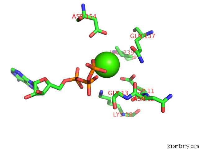
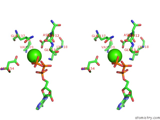
Calcium binding site 1 out of 4 in 2oan
Go back to
Calcium binding site 1 out
of 4 in the Structure of Oxidized Beta-Actin
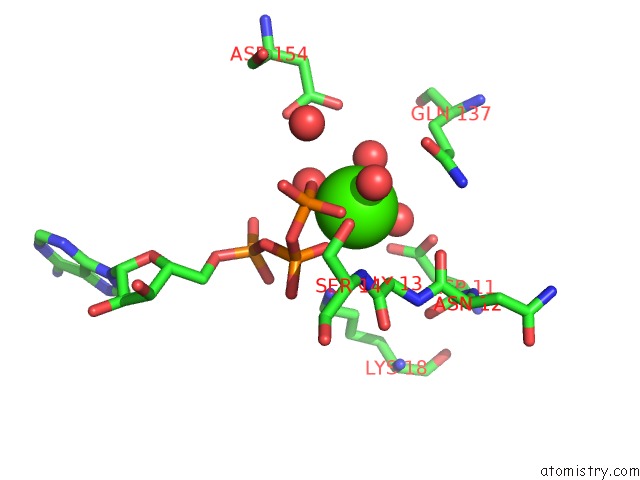
Mono view
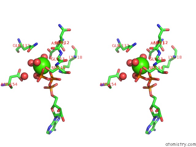
Stereo pair view
Mono view
Stereo pair view
A full contact list of Calcium with other atoms in the Ca binding
site number 1 of Structure of Oxidized Beta-Actin within 5.0Å range:
|
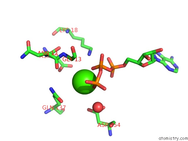
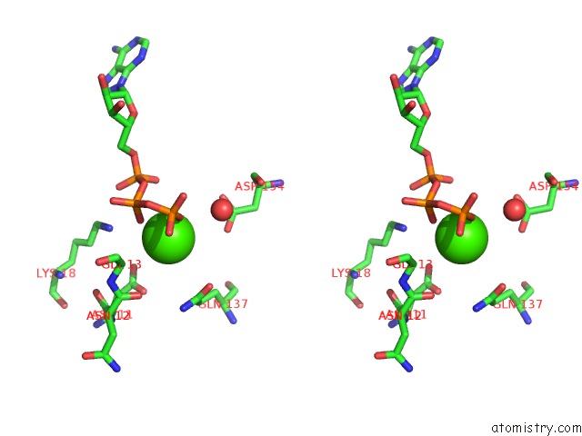
Calcium binding site 2 out of 4 in 2oan
Go back to
Calcium binding site 2 out
of 4 in the Structure of Oxidized Beta-Actin
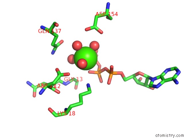
Mono view
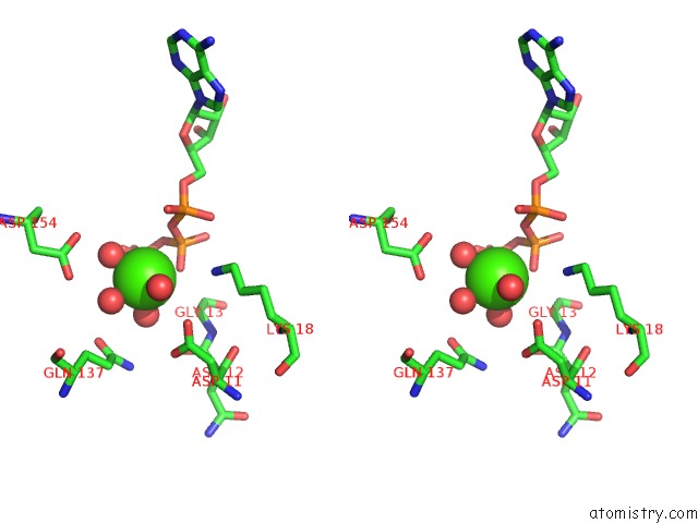
Stereo pair view
Mono view
Stereo pair view
A full contact list of Calcium with other atoms in the Ca binding
site number 2 of Structure of Oxidized Beta-Actin within 5.0Å range:
|
Calcium binding site 3 out of 4 in 2oan
Go back to
Calcium binding site 3 out
of 4 in the Structure of Oxidized Beta-Actin
Mono view
Stereo pair view
Mono view
Stereo pair view
A full contact list of Calcium with other atoms in the Ca binding
site number 3 of Structure of Oxidized Beta-Actin within 5.0Å range:
|
Calcium binding site 4 out of 4 in 2oan
Go back to
Calcium binding site 4 out
of 4 in the Structure of Oxidized Beta-Actin
Mono view
Stereo pair view
Mono view
Stereo pair view
A full contact list of Calcium with other atoms in the Ca binding
site number 4 of Structure of Oxidized Beta-Actin within 5.0Å range:
|
Reference:
I.Lassing,
F.Schmitzberger,
M.Bjornstedt,
A.Holmgren,
P.Nordlund,
C.E.Schutt,
U.Lindberg.
Molecular and Structural Basis For Redox Regulation of Beta-Actin. J.Mol.Biol. V. 370 331 2007.
ISSN: ISSN 0022-2836
PubMed: 17521670
DOI: 10.1016/J.JMB.2007.04.056
Page generated: Tue Jul 8 07:19:08 2025
ISSN: ISSN 0022-2836
PubMed: 17521670
DOI: 10.1016/J.JMB.2007.04.056
Last articles
Mg in 6C4CMg in 6C6K
Mg in 6C6O
Mg in 6C4A
Mg in 6C62
Mg in 6C5U
Mg in 6C5N
Mg in 6C55
Mg in 6C2W
Mg in 6C3P