Calcium »
PDB 2o8o-2ovu »
2oeo »
Calcium in PDB 2oeo: Cryogenic Crystal Structure of Staphylococcal Nuclease Variant Truncated Delta+Phs I92D
Enzymatic activity of Cryogenic Crystal Structure of Staphylococcal Nuclease Variant Truncated Delta+Phs I92D
All present enzymatic activity of Cryogenic Crystal Structure of Staphylococcal Nuclease Variant Truncated Delta+Phs I92D:
3.1.31.1;
3.1.31.1;
Protein crystallography data
The structure of Cryogenic Crystal Structure of Staphylococcal Nuclease Variant Truncated Delta+Phs I92D, PDB code: 2oeo
was solved by
R.L.Reynald,
E.E.Lattman,
A.G.Gittis,
with X-Ray Crystallography technique. A brief refinement statistics is given in the table below:
| Resolution Low / High (Å) | 20.79 / 2.00 |
| Space group | P 1 21 1 |
| Cell size a, b, c (Å), α, β, γ (°) | 30.452, 57.630, 34.618, 90.00, 99.74, 90.00 |
| R / Rfree (%) | 19.6 / 22.6 |
Calcium Binding Sites:
The binding sites of Calcium atom in the Cryogenic Crystal Structure of Staphylococcal Nuclease Variant Truncated Delta+Phs I92D
(pdb code 2oeo). This binding sites where shown within
5.0 Angstroms radius around Calcium atom.
In total only one binding site of Calcium was determined in the Cryogenic Crystal Structure of Staphylococcal Nuclease Variant Truncated Delta+Phs I92D, PDB code: 2oeo:
In total only one binding site of Calcium was determined in the Cryogenic Crystal Structure of Staphylococcal Nuclease Variant Truncated Delta+Phs I92D, PDB code: 2oeo:
Calcium binding site 1 out of 1 in 2oeo
Go back to
Calcium binding site 1 out
of 1 in the Cryogenic Crystal Structure of Staphylococcal Nuclease Variant Truncated Delta+Phs I92D
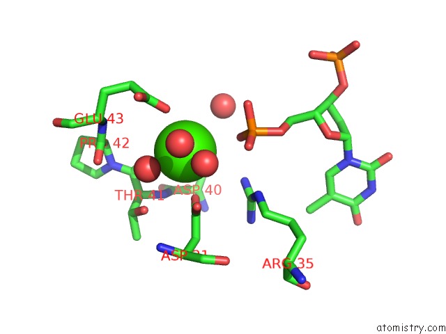
Mono view
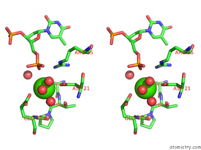
Stereo pair view
Mono view
Stereo pair view
A full contact list of Calcium with other atoms in the Ca binding
site number 1 of Cryogenic Crystal Structure of Staphylococcal Nuclease Variant Truncated Delta+Phs I92D within 5.0Å range:
|
Reference:
R.L.Reynald,
R.K.Gitti,
E.E.Lattman,
A.G.Gittis.
Buried Charges and Water in the Protein Interior: Reality or Fiction? To Be Published.
Page generated: Tue Jul 8 07:20:00 2025
Last articles
Mg in 6C6TMg in 6C6S
Mg in 6C4C
Mg in 6C6K
Mg in 6C6O
Mg in 6C4A
Mg in 6C62
Mg in 6C5U
Mg in 6C5N
Mg in 6C55