Calcium »
PDB 2o8o-2ovu »
2olg »
Calcium in PDB 2olg: Crystal Structure of the Serine Protease Domain of Prophenoloxidase Activating Factor-I in A Zymogen Form
Protein crystallography data
The structure of Crystal Structure of the Serine Protease Domain of Prophenoloxidase Activating Factor-I in A Zymogen Form, PDB code: 2olg
was solved by
N.C.Ha,
S.Piao,
with X-Ray Crystallography technique. A brief refinement statistics is given in the table below:
| Resolution Low / High (Å) | 27.42 / 1.70 |
| Space group | P 21 21 21 |
| Cell size a, b, c (Å), α, β, γ (°) | 38.229, 53.304, 116.643, 90.00, 90.00, 90.00 |
| R / Rfree (%) | 20.4 / 24.5 |
Calcium Binding Sites:
The binding sites of Calcium atom in the Crystal Structure of the Serine Protease Domain of Prophenoloxidase Activating Factor-I in A Zymogen Form
(pdb code 2olg). This binding sites where shown within
5.0 Angstroms radius around Calcium atom.
In total only one binding site of Calcium was determined in the Crystal Structure of the Serine Protease Domain of Prophenoloxidase Activating Factor-I in A Zymogen Form, PDB code: 2olg:
In total only one binding site of Calcium was determined in the Crystal Structure of the Serine Protease Domain of Prophenoloxidase Activating Factor-I in A Zymogen Form, PDB code: 2olg:
Calcium binding site 1 out of 1 in 2olg
Go back to
Calcium binding site 1 out
of 1 in the Crystal Structure of the Serine Protease Domain of Prophenoloxidase Activating Factor-I in A Zymogen Form
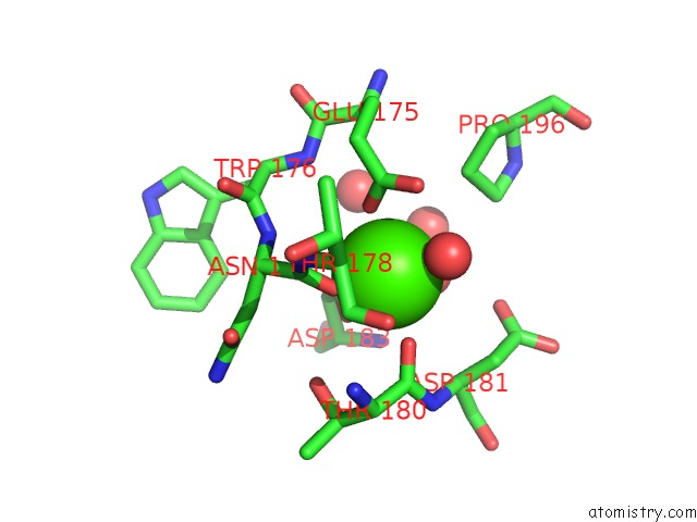
Mono view
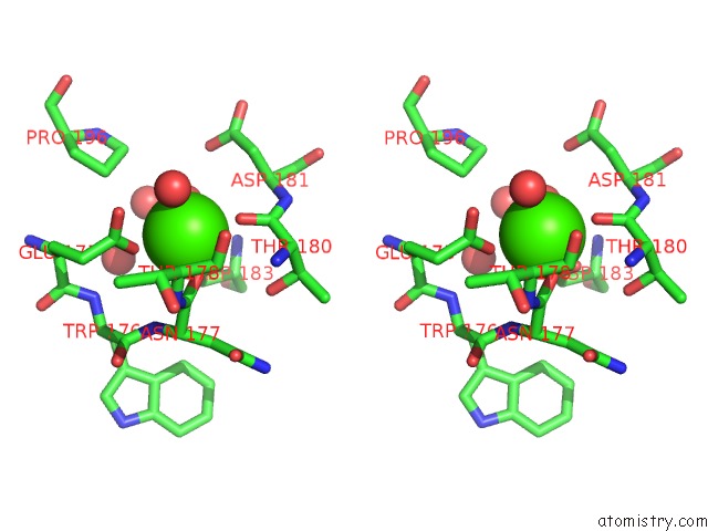
Stereo pair view
Mono view
Stereo pair view
A full contact list of Calcium with other atoms in the Ca binding
site number 1 of Crystal Structure of the Serine Protease Domain of Prophenoloxidase Activating Factor-I in A Zymogen Form within 5.0Å range:
|
Reference:
S.Piao,
S.Kim,
J.H.Kim,
J.W.Park,
B.L.Lee,
N.C.Ha.
Crystal Structure of the Serine Protease Domain of Prophenoloxidase Activating Factor-I J.Biol.Chem. V. 282 10783 2007.
ISSN: ISSN 0021-9258
PubMed: 17287215
DOI: 10.1074/JBC.M611556200
Page generated: Tue Jul 8 07:20:50 2025
ISSN: ISSN 0021-9258
PubMed: 17287215
DOI: 10.1074/JBC.M611556200
Last articles
Mg in 6C6SMg in 6C4C
Mg in 6C6K
Mg in 6C6O
Mg in 6C4A
Mg in 6C62
Mg in 6C5U
Mg in 6C5N
Mg in 6C55
Mg in 6C2W