Calcium »
PDB 2o8o-2ovu »
2ost »
Calcium in PDB 2ost: The Structure of A Bacterial Homing Endonuclease : I-SSP6803I
Protein crystallography data
The structure of The Structure of A Bacterial Homing Endonuclease : I-SSP6803I, PDB code: 2ost
was solved by
L.Zhao,
R.P.Bonocora,
D.A.Shub,
B.L.Stoddard,
with X-Ray Crystallography technique. A brief refinement statistics is given in the table below:
| Resolution Low / High (Å) | 160.13 / 3.10 |
| Space group | I 4 2 2 |
| Cell size a, b, c (Å), α, β, γ (°) | 143.777, 143.777, 319.179, 90.00, 90.00, 90.00 |
| R / Rfree (%) | 27.8 / 32.8 |
Calcium Binding Sites:
The binding sites of Calcium atom in the The Structure of A Bacterial Homing Endonuclease : I-SSP6803I
(pdb code 2ost). This binding sites where shown within
5.0 Angstroms radius around Calcium atom.
In total 2 binding sites of Calcium where determined in the The Structure of A Bacterial Homing Endonuclease : I-SSP6803I, PDB code: 2ost:
Jump to Calcium binding site number: 1; 2;
In total 2 binding sites of Calcium where determined in the The Structure of A Bacterial Homing Endonuclease : I-SSP6803I, PDB code: 2ost:
Jump to Calcium binding site number: 1; 2;
Calcium binding site 1 out of 2 in 2ost
Go back to
Calcium binding site 1 out
of 2 in the The Structure of A Bacterial Homing Endonuclease : I-SSP6803I
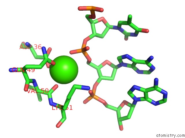
Mono view
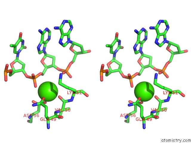
Stereo pair view
Mono view
Stereo pair view
A full contact list of Calcium with other atoms in the Ca binding
site number 1 of The Structure of A Bacterial Homing Endonuclease : I-SSP6803I within 5.0Å range:
|
Calcium binding site 2 out of 2 in 2ost
Go back to
Calcium binding site 2 out
of 2 in the The Structure of A Bacterial Homing Endonuclease : I-SSP6803I
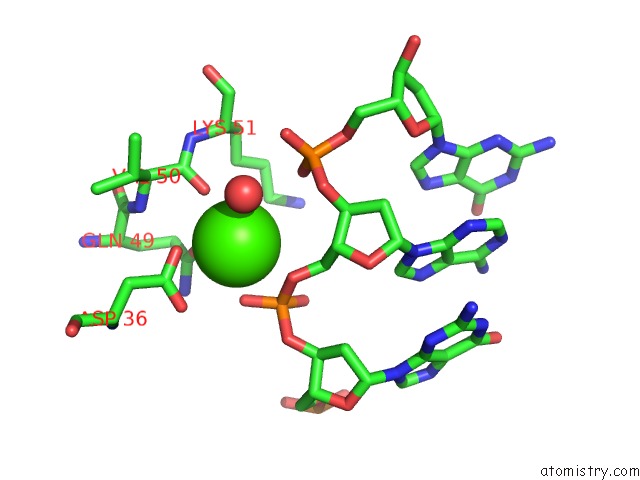
Mono view
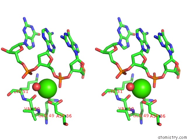
Stereo pair view
Mono view
Stereo pair view
A full contact list of Calcium with other atoms in the Ca binding
site number 2 of The Structure of A Bacterial Homing Endonuclease : I-SSP6803I within 5.0Å range:
|
Reference:
L.Zhao,
R.P.Bonocora,
D.A.Shub,
B.L.Stoddard.
The Restriction Fold Turns to the Dark Side: A Bacterial Homing Endonuclease with A Pd-(D/E)-Xk Motif. Embo J. V. 26 2432 2007.
ISSN: ISSN 0261-4189
PubMed: 17410205
DOI: 10.1038/SJ.EMBOJ.7601672
Page generated: Tue Jul 8 07:24:40 2025
ISSN: ISSN 0261-4189
PubMed: 17410205
DOI: 10.1038/SJ.EMBOJ.7601672
Last articles
Mg in 6FCBMg in 6FBV
Mg in 6FBP
Mg in 6FBO
Mg in 6FBN
Mg in 6FBE
Mg in 6FBD
Mg in 6FBC
Mg in 6FBB
Mg in 6FB8