Calcium »
PDB 2ovx-2p9k »
2owl »
Calcium in PDB 2owl: Crystal Structure of E. Coli Rdgc
Protein crystallography data
The structure of Crystal Structure of E. Coli Rdgc, PDB code: 2owl
was solved by
G.S.Briggs,
P.A.Mcewan,
J.Yu,
T.Moore,
J.Emsley,
R.G.Lloyd,
with X-Ray Crystallography technique. A brief refinement statistics is given in the table below:
| Resolution Low / High (Å) | 19.80 / 2.40 |
| Space group | P 21 21 21 |
| Cell size a, b, c (Å), α, β, γ (°) | 51.425, 95.683, 171.576, 90.00, 90.00, 90.00 |
| R / Rfree (%) | 23.2 / 32.6 |
Calcium Binding Sites:
The binding sites of Calcium atom in the Crystal Structure of E. Coli Rdgc
(pdb code 2owl). This binding sites where shown within
5.0 Angstroms radius around Calcium atom.
In total 2 binding sites of Calcium where determined in the Crystal Structure of E. Coli Rdgc, PDB code: 2owl:
Jump to Calcium binding site number: 1; 2;
In total 2 binding sites of Calcium where determined in the Crystal Structure of E. Coli Rdgc, PDB code: 2owl:
Jump to Calcium binding site number: 1; 2;
Calcium binding site 1 out of 2 in 2owl
Go back to
Calcium binding site 1 out
of 2 in the Crystal Structure of E. Coli Rdgc
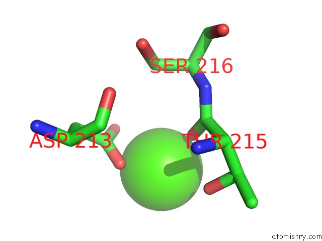
Mono view
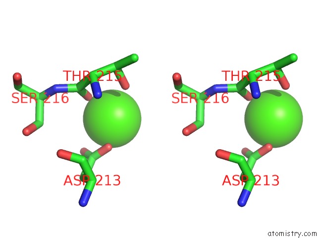
Stereo pair view
Mono view
Stereo pair view
A full contact list of Calcium with other atoms in the Ca binding
site number 1 of Crystal Structure of E. Coli Rdgc within 5.0Å range:
|
Calcium binding site 2 out of 2 in 2owl
Go back to
Calcium binding site 2 out
of 2 in the Crystal Structure of E. Coli Rdgc
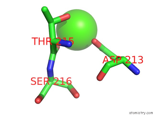
Mono view
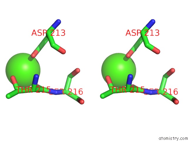
Stereo pair view
Mono view
Stereo pair view
A full contact list of Calcium with other atoms in the Ca binding
site number 2 of Crystal Structure of E. Coli Rdgc within 5.0Å range:
|
Reference:
G.S.Briggs,
P.A.Mcewan,
J.Yu,
T.Moore,
J.Emsley,
R.G.Lloyd.
Ring Structure of the Escherichia Coli Dna-Binding Protein Rdgc Associated with Recombination and Replication Fork Repair. J.Biol.Chem. V. 282 12353 2007.
ISSN: ISSN 0021-9258
PubMed: 17308310
DOI: 10.1074/JBC.C700023200
Page generated: Tue Jul 8 07:28:31 2025
ISSN: ISSN 0021-9258
PubMed: 17308310
DOI: 10.1074/JBC.C700023200
Last articles
Mg in 8CGIMg in 8CF8
Mg in 8CGA
Mg in 8CEP
Mg in 8CF1
Mg in 8CAH
Mg in 8CE5
Mg in 8CDQ
Mg in 8CE2
Mg in 8CCO