Calcium »
PDB 2ovx-2p9k »
2oyh »
Calcium in PDB 2oyh: Crystal Structure of Fragment D of GAMMAD298,301A Fibrinogen with the Peptide Ligand Gly-His-Arg-Pro-Amide
Protein crystallography data
The structure of Crystal Structure of Fragment D of GAMMAD298,301A Fibrinogen with the Peptide Ligand Gly-His-Arg-Pro-Amide, PDB code: 2oyh
was solved by
M.S.Kostelansky,
O.V.Gorkun,
S.T.Lord,
with X-Ray Crystallography technique. A brief refinement statistics is given in the table below:
| Resolution Low / High (Å) | 18.00 / 2.40 |
| Space group | P 21 21 21 |
| Cell size a, b, c (Å), α, β, γ (°) | 89.164, 94.119, 226.730, 90.00, 90.00, 90.00 |
| R / Rfree (%) | 22 / 25.9 |
Calcium Binding Sites:
The binding sites of Calcium atom in the Crystal Structure of Fragment D of GAMMAD298,301A Fibrinogen with the Peptide Ligand Gly-His-Arg-Pro-Amide
(pdb code 2oyh). This binding sites where shown within
5.0 Angstroms radius around Calcium atom.
In total 4 binding sites of Calcium where determined in the Crystal Structure of Fragment D of GAMMAD298,301A Fibrinogen with the Peptide Ligand Gly-His-Arg-Pro-Amide, PDB code: 2oyh:
Jump to Calcium binding site number: 1; 2; 3; 4;
In total 4 binding sites of Calcium where determined in the Crystal Structure of Fragment D of GAMMAD298,301A Fibrinogen with the Peptide Ligand Gly-His-Arg-Pro-Amide, PDB code: 2oyh:
Jump to Calcium binding site number: 1; 2; 3; 4;
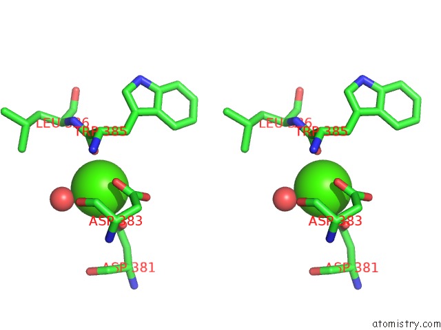
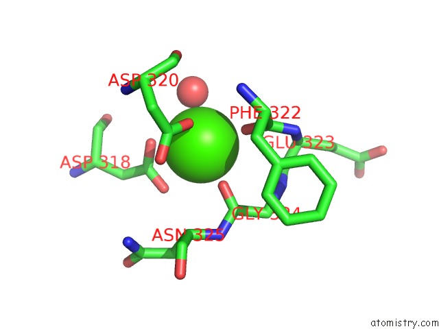
Calcium binding site 1 out of 4 in 2oyh
Go back to
Calcium binding site 1 out
of 4 in the Crystal Structure of Fragment D of GAMMAD298,301A Fibrinogen with the Peptide Ligand Gly-His-Arg-Pro-Amide

Mono view
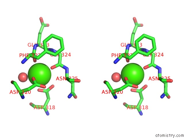
Stereo pair view
Mono view
Stereo pair view
A full contact list of Calcium with other atoms in the Ca binding
site number 1 of Crystal Structure of Fragment D of GAMMAD298,301A Fibrinogen with the Peptide Ligand Gly-His-Arg-Pro-Amide within 5.0Å range:
|
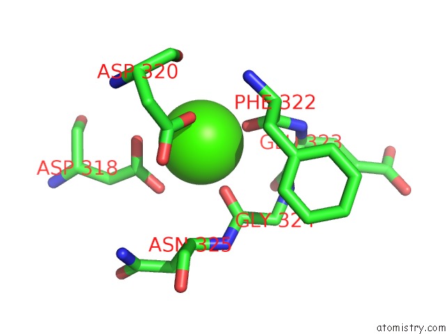
Calcium binding site 2 out of 4 in 2oyh
Go back to
Calcium binding site 2 out
of 4 in the Crystal Structure of Fragment D of GAMMAD298,301A Fibrinogen with the Peptide Ligand Gly-His-Arg-Pro-Amide
Mono view
Stereo pair view
Mono view
Stereo pair view
A full contact list of Calcium with other atoms in the Ca binding
site number 2 of Crystal Structure of Fragment D of GAMMAD298,301A Fibrinogen with the Peptide Ligand Gly-His-Arg-Pro-Amide within 5.0Å range:
|
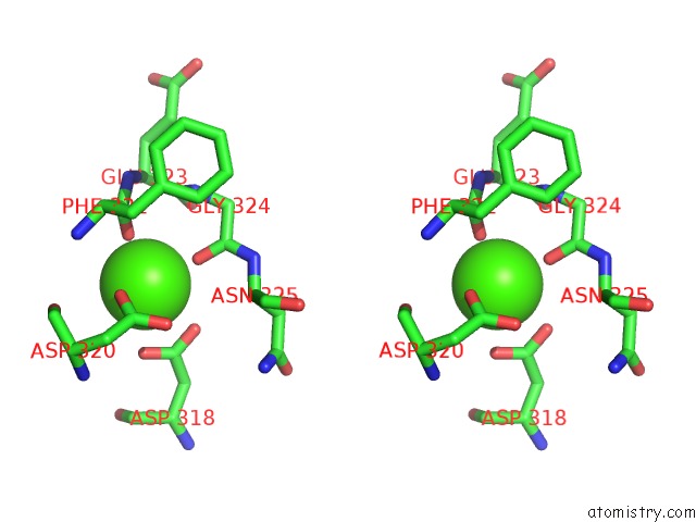
Calcium binding site 3 out of 4 in 2oyh
Go back to
Calcium binding site 3 out
of 4 in the Crystal Structure of Fragment D of GAMMAD298,301A Fibrinogen with the Peptide Ligand Gly-His-Arg-Pro-Amide

Mono view
Stereo pair view
Mono view
Stereo pair view
A full contact list of Calcium with other atoms in the Ca binding
site number 3 of Crystal Structure of Fragment D of GAMMAD298,301A Fibrinogen with the Peptide Ligand Gly-His-Arg-Pro-Amide within 5.0Å range:
|
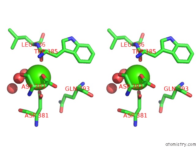
Calcium binding site 4 out of 4 in 2oyh
Go back to
Calcium binding site 4 out
of 4 in the Crystal Structure of Fragment D of GAMMAD298,301A Fibrinogen with the Peptide Ligand Gly-His-Arg-Pro-Amide
Mono view
Stereo pair view
Mono view
Stereo pair view
A full contact list of Calcium with other atoms in the Ca binding
site number 4 of Crystal Structure of Fragment D of GAMMAD298,301A Fibrinogen with the Peptide Ligand Gly-His-Arg-Pro-Amide within 5.0Å range:
|
Reference:
M.S.Kostelansky,
K.C.Lounes,
L.F.Ping,
S.K.Dickerson,
O.V.Gorkun,
S.T.Lord.
Probing the GAMMA2 Calcium-Binding Site: Studies with GAMMAD298,301A Fibrinogen Reveal Changes in the GAMMA294-301 Loop That Alter the Integrity of the "A" Polymerization Site. Biochemistry V. 46 5114 2007.
ISSN: ISSN 0006-2960
PubMed: 17411074
DOI: 10.1021/BI602607A
Page generated: Tue Jul 8 07:31:29 2025
ISSN: ISSN 0006-2960
PubMed: 17411074
DOI: 10.1021/BI602607A
Last articles
Mg in 8EZBMg in 8EZF
Mg in 8EYE
Mg in 8EZ2
Mg in 8EXX
Mg in 8EX3
Mg in 8EXY
Mg in 8EVZ
Mg in 8EXW
Mg in 8EWZ