Calcium »
PDB 2p9n-2prk »
2pc6 »
Calcium in PDB 2pc6: Crystal Structure of Putative Acetolactate Synthase- Small Subunit From Nitrosomonas Europaea
Enzymatic activity of Crystal Structure of Putative Acetolactate Synthase- Small Subunit From Nitrosomonas Europaea
All present enzymatic activity of Crystal Structure of Putative Acetolactate Synthase- Small Subunit From Nitrosomonas Europaea:
4.1.3.18;
4.1.3.18;
Protein crystallography data
The structure of Crystal Structure of Putative Acetolactate Synthase- Small Subunit From Nitrosomonas Europaea, PDB code: 2pc6
was solved by
J.J.Petkowski,
M.Chruszcz,
M.D.Zimmerman,
H.Zheng,
M.T.Cymborowski,
T.Skarina,
O.Onopriyenko,
A.Savchenko,
A.Edwards,
W.Minor,
A.Joachimiak,
Midwest Center For Structural Genomics (Mcsg),
with X-Ray Crystallography technique. A brief refinement statistics is given in the table below:
| Resolution Low / High (Å) | 35.58 / 2.50 |
| Space group | P 42 21 2 |
| Cell size a, b, c (Å), α, β, γ (°) | 122.123, 122.123, 111.559, 90.00, 90.00, 90.00 |
| R / Rfree (%) | 20.5 / 27.6 |
Calcium Binding Sites:
The binding sites of Calcium atom in the Crystal Structure of Putative Acetolactate Synthase- Small Subunit From Nitrosomonas Europaea
(pdb code 2pc6). This binding sites where shown within
5.0 Angstroms radius around Calcium atom.
In total 3 binding sites of Calcium where determined in the Crystal Structure of Putative Acetolactate Synthase- Small Subunit From Nitrosomonas Europaea, PDB code: 2pc6:
Jump to Calcium binding site number: 1; 2; 3;
In total 3 binding sites of Calcium where determined in the Crystal Structure of Putative Acetolactate Synthase- Small Subunit From Nitrosomonas Europaea, PDB code: 2pc6:
Jump to Calcium binding site number: 1; 2; 3;
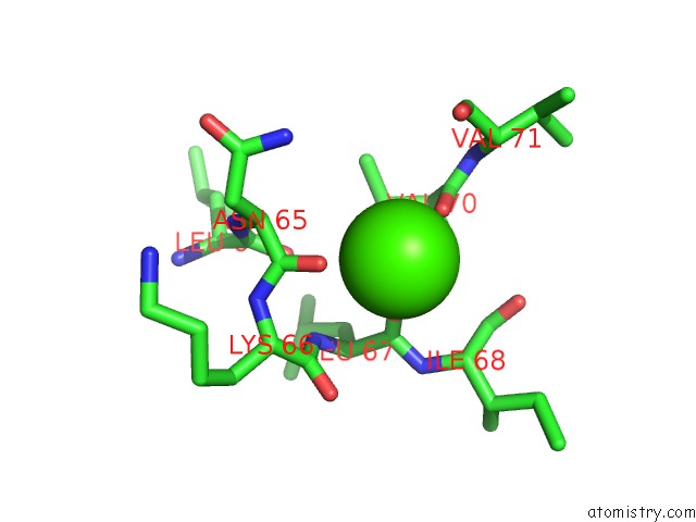
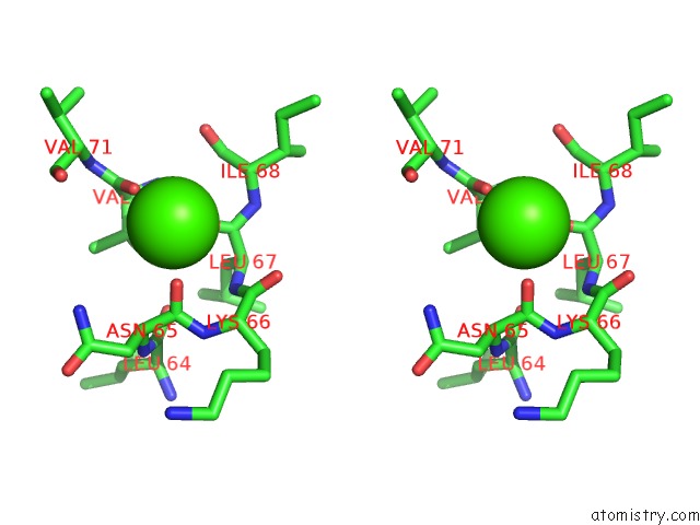
Calcium binding site 1 out of 3 in 2pc6
Go back to
Calcium binding site 1 out
of 3 in the Crystal Structure of Putative Acetolactate Synthase- Small Subunit From Nitrosomonas Europaea
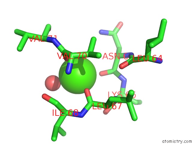
Mono view
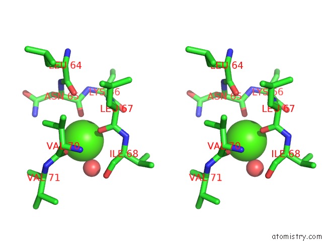
Stereo pair view
Mono view
Stereo pair view
A full contact list of Calcium with other atoms in the Ca binding
site number 1 of Crystal Structure of Putative Acetolactate Synthase- Small Subunit From Nitrosomonas Europaea within 5.0Å range:
|
Calcium binding site 2 out of 3 in 2pc6
Go back to
Calcium binding site 2 out
of 3 in the Crystal Structure of Putative Acetolactate Synthase- Small Subunit From Nitrosomonas Europaea
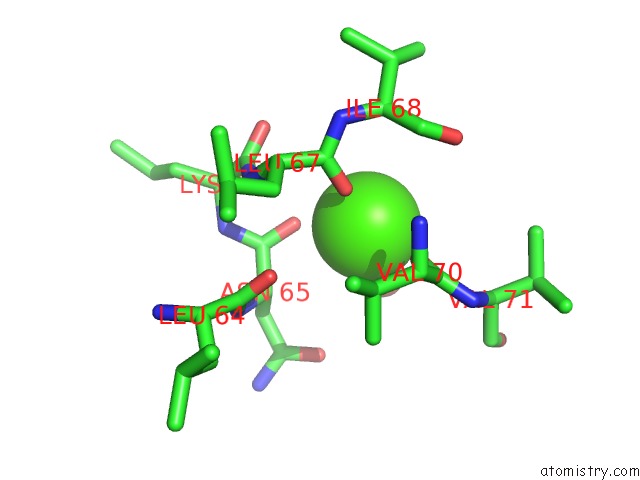
Mono view
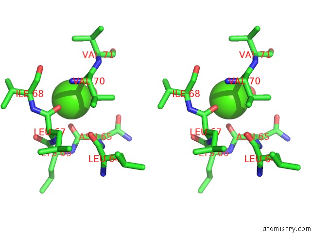
Stereo pair view
Mono view
Stereo pair view
A full contact list of Calcium with other atoms in the Ca binding
site number 2 of Crystal Structure of Putative Acetolactate Synthase- Small Subunit From Nitrosomonas Europaea within 5.0Å range:
|
Calcium binding site 3 out of 3 in 2pc6
Go back to
Calcium binding site 3 out
of 3 in the Crystal Structure of Putative Acetolactate Synthase- Small Subunit From Nitrosomonas Europaea
Mono view
Stereo pair view
Mono view
Stereo pair view
A full contact list of Calcium with other atoms in the Ca binding
site number 3 of Crystal Structure of Putative Acetolactate Synthase- Small Subunit From Nitrosomonas Europaea within 5.0Å range:
|
Reference:
J.J.Petkowski,
M.Chruszcz,
M.D.Zimmerman,
H.Zheng,
T.Skarina,
O.Onopriyenko,
M.T.Cymborowski,
K.D.Koclega,
A.Savchenko,
A.Edwards,
W.Minor.
Crystal Structures of TM0549 and NE1324--Two Orthologs of E. Coli Ahas Isozyme III Small Regulatory Subunit. Protein Sci. V. 16 1360 2007.
ISSN: ISSN 0961-8368
PubMed: 17586771
DOI: 10.1110/PS.072793807
Page generated: Tue Jul 8 07:40:19 2025
ISSN: ISSN 0961-8368
PubMed: 17586771
DOI: 10.1110/PS.072793807
Last articles
K in 6AI6K in 6AU4
K in 6ASO
K in 6AFZ
K in 6AFY
K in 6AFX
K in 6AFW
K in 6AFV
K in 6AFU
K in 6AFT