Calcium »
PDB 2p9n-2prk »
2pf2 »
Calcium in PDB 2pf2: The Ca+2 Ion and Membrane Binding Structure of the Gla Domain of Ca- Prothrombin Fragment 1
Protein crystallography data
The structure of The Ca+2 Ion and Membrane Binding Structure of the Gla Domain of Ca- Prothrombin Fragment 1, PDB code: 2pf2
was solved by
M.Soriano-Garcia,
K.Padmanabhan,
A.M.De Vos,
A.Tulinsky,
with X-Ray Crystallography technique. A brief refinement statistics is given in the table below:
| Resolution Low / High (Å) | 7.00 / 2.20 |
| Space group | P 21 21 21 |
| Cell size a, b, c (Å), α, β, γ (°) | 39.390, 53.880, 129.640, 90.00, 90.00, 90.00 |
| R / Rfree (%) | n/a / n/a |
Calcium Binding Sites:
The binding sites of Calcium atom in the The Ca+2 Ion and Membrane Binding Structure of the Gla Domain of Ca- Prothrombin Fragment 1
(pdb code 2pf2). This binding sites where shown within
5.0 Angstroms radius around Calcium atom.
In total 7 binding sites of Calcium where determined in the The Ca+2 Ion and Membrane Binding Structure of the Gla Domain of Ca- Prothrombin Fragment 1, PDB code: 2pf2:
Jump to Calcium binding site number: 1; 2; 3; 4; 5; 6; 7;
In total 7 binding sites of Calcium where determined in the The Ca+2 Ion and Membrane Binding Structure of the Gla Domain of Ca- Prothrombin Fragment 1, PDB code: 2pf2:
Jump to Calcium binding site number: 1; 2; 3; 4; 5; 6; 7;
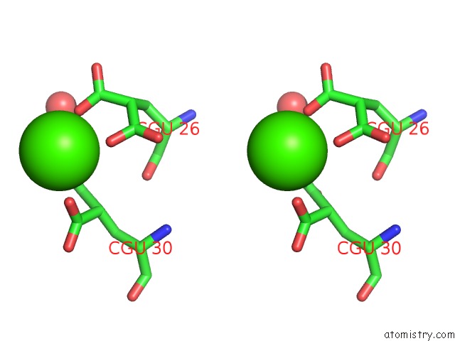
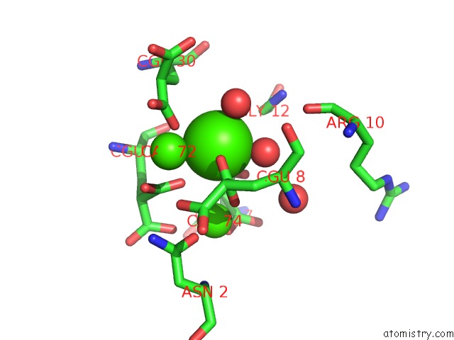
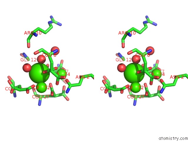
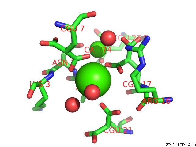
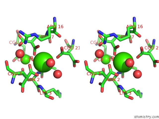
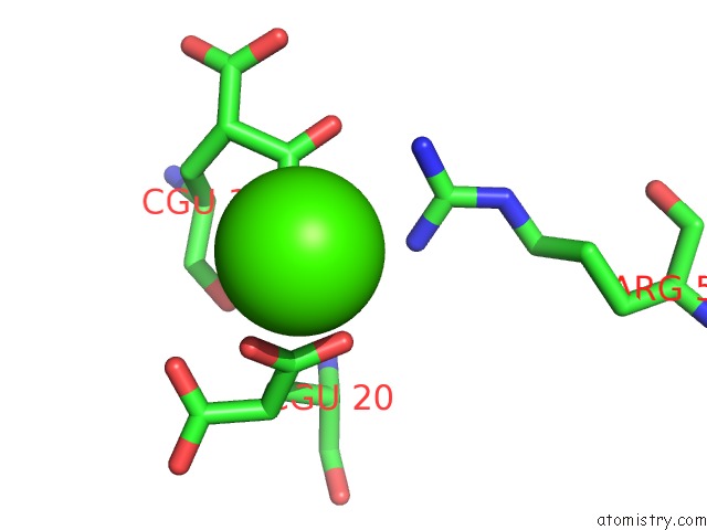
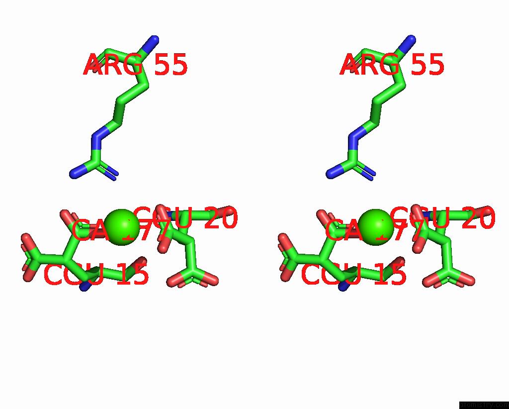
Calcium binding site 1 out of 7 in 2pf2
Go back to
Calcium binding site 1 out
of 7 in the The Ca+2 Ion and Membrane Binding Structure of the Gla Domain of Ca- Prothrombin Fragment 1
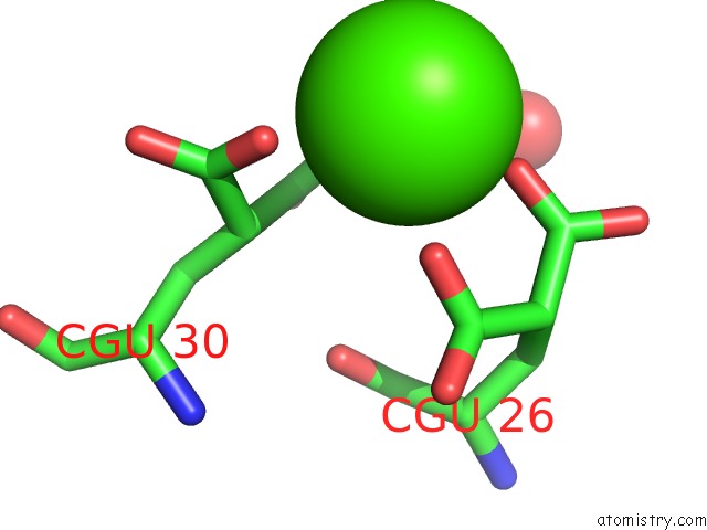
Mono view
Stereo pair view
Mono view
Stereo pair view
A full contact list of Calcium with other atoms in the Ca binding
site number 1 of The Ca+2 Ion and Membrane Binding Structure of the Gla Domain of Ca- Prothrombin Fragment 1 within 5.0Å range:
|
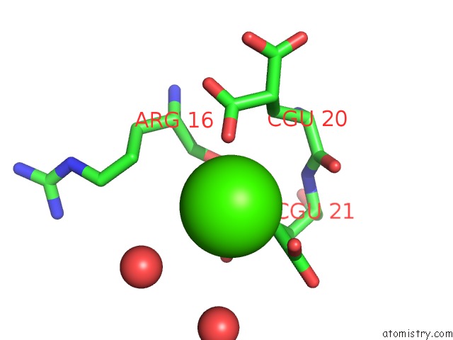
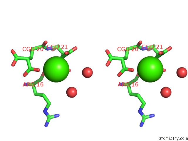
Calcium binding site 2 out of 7 in 2pf2
Go back to
Calcium binding site 2 out
of 7 in the The Ca+2 Ion and Membrane Binding Structure of the Gla Domain of Ca- Prothrombin Fragment 1
Mono view
Stereo pair view
Mono view
Stereo pair view
A full contact list of Calcium with other atoms in the Ca binding
site number 2 of The Ca+2 Ion and Membrane Binding Structure of the Gla Domain of Ca- Prothrombin Fragment 1 within 5.0Å range:
|
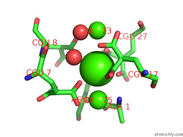
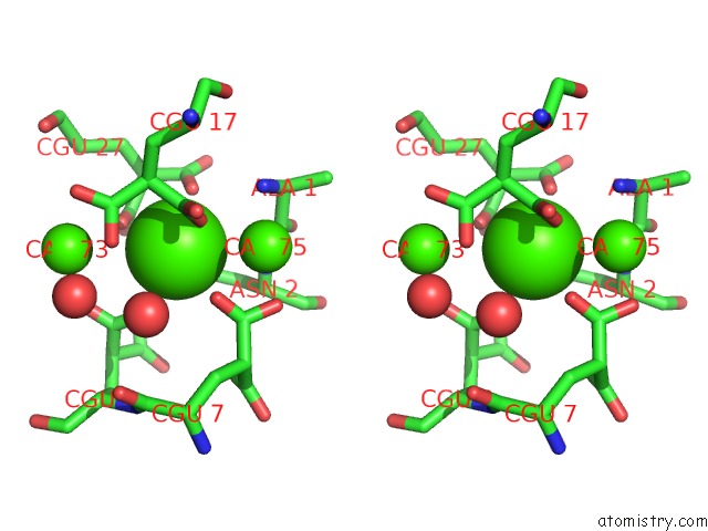
Calcium binding site 3 out of 7 in 2pf2
Go back to
Calcium binding site 3 out
of 7 in the The Ca+2 Ion and Membrane Binding Structure of the Gla Domain of Ca- Prothrombin Fragment 1
Mono view
Stereo pair view
Mono view
Stereo pair view
A full contact list of Calcium with other atoms in the Ca binding
site number 3 of The Ca+2 Ion and Membrane Binding Structure of the Gla Domain of Ca- Prothrombin Fragment 1 within 5.0Å range:
|
Calcium binding site 4 out of 7 in 2pf2
Go back to
Calcium binding site 4 out
of 7 in the The Ca+2 Ion and Membrane Binding Structure of the Gla Domain of Ca- Prothrombin Fragment 1
Mono view
Stereo pair view
Mono view
Stereo pair view
A full contact list of Calcium with other atoms in the Ca binding
site number 4 of The Ca+2 Ion and Membrane Binding Structure of the Gla Domain of Ca- Prothrombin Fragment 1 within 5.0Å range:
|
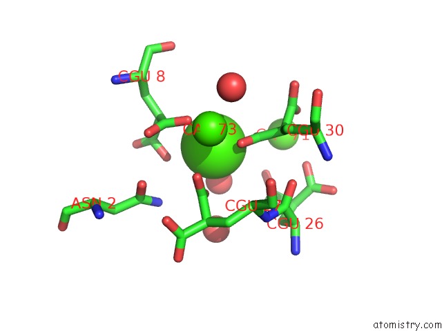
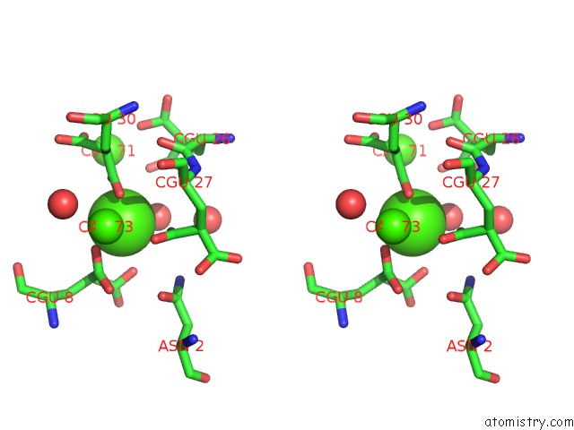
Calcium binding site 5 out of 7 in 2pf2
Go back to
Calcium binding site 5 out
of 7 in the The Ca+2 Ion and Membrane Binding Structure of the Gla Domain of Ca- Prothrombin Fragment 1
Mono view
Stereo pair view
Mono view
Stereo pair view
A full contact list of Calcium with other atoms in the Ca binding
site number 5 of The Ca+2 Ion and Membrane Binding Structure of the Gla Domain of Ca- Prothrombin Fragment 1 within 5.0Å range:
|
Calcium binding site 6 out of 7 in 2pf2
Go back to
Calcium binding site 6 out
of 7 in the The Ca+2 Ion and Membrane Binding Structure of the Gla Domain of Ca- Prothrombin Fragment 1
Mono view
Stereo pair view
Mono view
Stereo pair view
A full contact list of Calcium with other atoms in the Ca binding
site number 6 of The Ca+2 Ion and Membrane Binding Structure of the Gla Domain of Ca- Prothrombin Fragment 1 within 5.0Å range:
|
Calcium binding site 7 out of 7 in 2pf2
Go back to
Calcium binding site 7 out
of 7 in the The Ca+2 Ion and Membrane Binding Structure of the Gla Domain of Ca- Prothrombin Fragment 1
Mono view
Stereo pair view
Mono view
Stereo pair view
A full contact list of Calcium with other atoms in the Ca binding
site number 7 of The Ca+2 Ion and Membrane Binding Structure of the Gla Domain of Ca- Prothrombin Fragment 1 within 5.0Å range:
|
Reference:
M.Soriano-Garcia,
K.Padmanabhan,
A.M.De Vos,
A.Tulinsky.
The CA2+ Ion and Membrane Binding Structure of the Gla Domain of Ca-Prothrombin Fragment 1. Biochemistry V. 31 2554 1992.
ISSN: ISSN 0006-2960
PubMed: 1547238
DOI: 10.1021/BI00124A016
Page generated: Tue Jul 8 07:41:04 2025
ISSN: ISSN 0006-2960
PubMed: 1547238
DOI: 10.1021/BI00124A016
Last articles
K in 5UAMK in 5U3S
K in 5U41
K in 5U3Q
K in 5U3G
K in 5TZO
K in 5U06
K in 5TXG
K in 5TVV
K in 5TXR