Calcium »
PDB 2p9n-2prk »
2pq2 »
Calcium in PDB 2pq2: Structure of Serine Proteinase K Complex with A Highly Flexible Hydrophobic Peptide at 1.8A Resolution
Enzymatic activity of Structure of Serine Proteinase K Complex with A Highly Flexible Hydrophobic Peptide at 1.8A Resolution
All present enzymatic activity of Structure of Serine Proteinase K Complex with A Highly Flexible Hydrophobic Peptide at 1.8A Resolution:
3.4.21.64;
3.4.21.64;
Protein crystallography data
The structure of Structure of Serine Proteinase K Complex with A Highly Flexible Hydrophobic Peptide at 1.8A Resolution, PDB code: 2pq2
was solved by
A.S.Ethayathulla,
A.K.Singh,
N.Singh,
S.Sharma,
M.Sinha,
R.K.Somvanshi,
P.Kaur,
S.Dey,
A.Srinivasan,
T.P.Singh,
with X-Ray Crystallography technique. A brief refinement statistics is given in the table below:
| Resolution Low / High (Å) | 57.30 / 1.82 |
| Space group | P 43 21 2 |
| Cell size a, b, c (Å), α, β, γ (°) | 68.314, 68.314, 108.418, 90.00, 90.00, 90.00 |
| R / Rfree (%) | 16.8 / 20.7 |
Calcium Binding Sites:
The binding sites of Calcium atom in the Structure of Serine Proteinase K Complex with A Highly Flexible Hydrophobic Peptide at 1.8A Resolution
(pdb code 2pq2). This binding sites where shown within
5.0 Angstroms radius around Calcium atom.
In total only one binding site of Calcium was determined in the Structure of Serine Proteinase K Complex with A Highly Flexible Hydrophobic Peptide at 1.8A Resolution, PDB code: 2pq2:
In total only one binding site of Calcium was determined in the Structure of Serine Proteinase K Complex with A Highly Flexible Hydrophobic Peptide at 1.8A Resolution, PDB code: 2pq2:
Calcium binding site 1 out of 1 in 2pq2
Go back to
Calcium binding site 1 out
of 1 in the Structure of Serine Proteinase K Complex with A Highly Flexible Hydrophobic Peptide at 1.8A Resolution
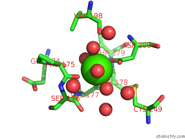
Mono view
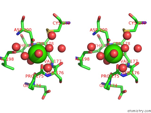
Stereo pair view
Mono view
Stereo pair view
A full contact list of Calcium with other atoms in the Ca binding
site number 1 of Structure of Serine Proteinase K Complex with A Highly Flexible Hydrophobic Peptide at 1.8A Resolution within 5.0Å range:
|
Reference:
A.S.Ethayathulla,
A.K.Singh,
N.Singh,
S.Sharma,
M.Sinha,
R.K.Somvanshi,
P.Kaur,
S.Dey,
A.Srinivasan,
T.P.Singh.
Structure of Serine Proteinase K Complex with A Highly Flexible Hydrophobic Peptide at 1.8A Resolution To Be Published.
Page generated: Tue Jul 8 07:45:56 2025
Last articles
K in 5SCKK in 5SCJ
K in 5SCI
K in 5SCH
K in 5SCG
K in 5SCF
K in 5SCE
K in 5SCD
K in 5SCA
K in 5SCB