Calcium »
PDB 2p9n-2prk »
2prk »
Calcium in PDB 2prk: Synchrotron X-Ray Data Collection and Restrained Least-Squares Refinement of the Crystal Structure of Proteinase K at 1.5 Angstroms Resolution
Enzymatic activity of Synchrotron X-Ray Data Collection and Restrained Least-Squares Refinement of the Crystal Structure of Proteinase K at 1.5 Angstroms Resolution
All present enzymatic activity of Synchrotron X-Ray Data Collection and Restrained Least-Squares Refinement of the Crystal Structure of Proteinase K at 1.5 Angstroms Resolution:
3.4.21.14;
3.4.21.14;
Protein crystallography data
The structure of Synchrotron X-Ray Data Collection and Restrained Least-Squares Refinement of the Crystal Structure of Proteinase K at 1.5 Angstroms Resolution, PDB code: 2prk
was solved by
C.Betzel,
G.P.Pal,
W.Saenger,
with X-Ray Crystallography technique. A brief refinement statistics is given in the table below:
| Resolution Low / High (Å) | N/A / 1.50 |
| Space group | P 43 21 2 |
| Cell size a, b, c (Å), α, β, γ (°) | 68.170, 68.170, 108.260, 90.00, 90.00, 90.00 |
| R / Rfree (%) | n/a / n/a |
Calcium Binding Sites:
The binding sites of Calcium atom in the Synchrotron X-Ray Data Collection and Restrained Least-Squares Refinement of the Crystal Structure of Proteinase K at 1.5 Angstroms Resolution
(pdb code 2prk). This binding sites where shown within
5.0 Angstroms radius around Calcium atom.
In total 2 binding sites of Calcium where determined in the Synchrotron X-Ray Data Collection and Restrained Least-Squares Refinement of the Crystal Structure of Proteinase K at 1.5 Angstroms Resolution, PDB code: 2prk:
Jump to Calcium binding site number: 1; 2;
In total 2 binding sites of Calcium where determined in the Synchrotron X-Ray Data Collection and Restrained Least-Squares Refinement of the Crystal Structure of Proteinase K at 1.5 Angstroms Resolution, PDB code: 2prk:
Jump to Calcium binding site number: 1; 2;
Calcium binding site 1 out of 2 in 2prk
Go back to
Calcium binding site 1 out
of 2 in the Synchrotron X-Ray Data Collection and Restrained Least-Squares Refinement of the Crystal Structure of Proteinase K at 1.5 Angstroms Resolution
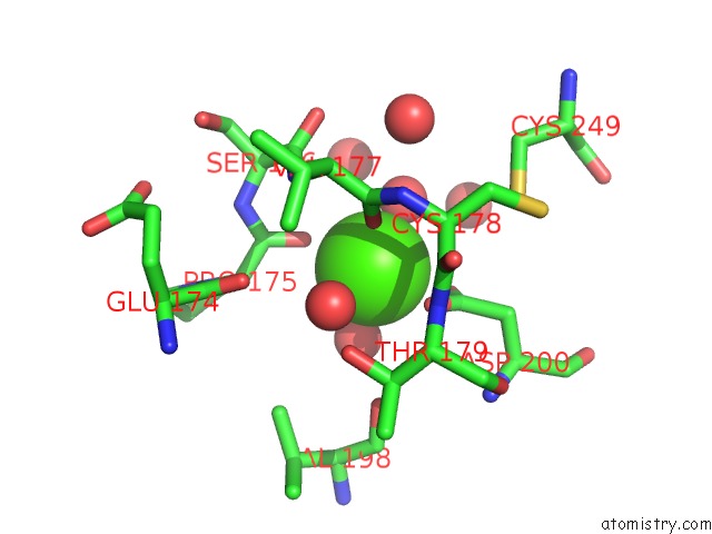
Mono view
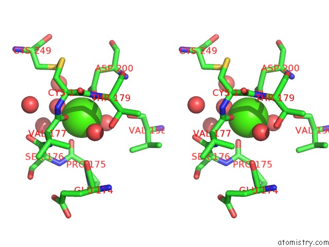
Stereo pair view
Mono view
Stereo pair view
A full contact list of Calcium with other atoms in the Ca binding
site number 1 of Synchrotron X-Ray Data Collection and Restrained Least-Squares Refinement of the Crystal Structure of Proteinase K at 1.5 Angstroms Resolution within 5.0Å range:
|
Calcium binding site 2 out of 2 in 2prk
Go back to
Calcium binding site 2 out
of 2 in the Synchrotron X-Ray Data Collection and Restrained Least-Squares Refinement of the Crystal Structure of Proteinase K at 1.5 Angstroms Resolution
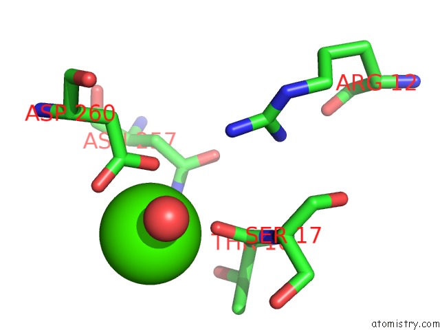
Mono view
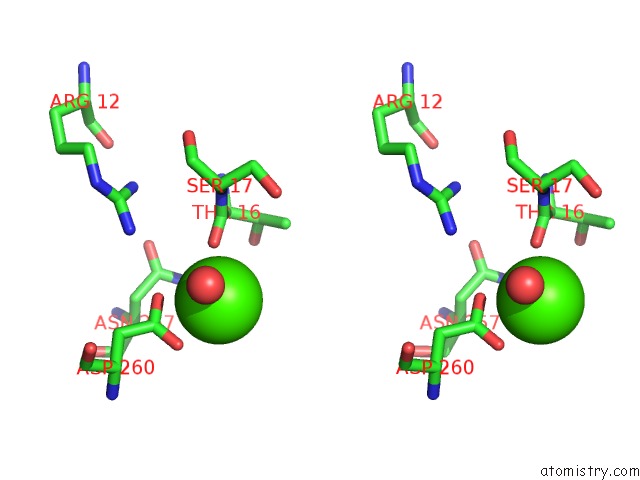
Stereo pair view
Mono view
Stereo pair view
A full contact list of Calcium with other atoms in the Ca binding
site number 2 of Synchrotron X-Ray Data Collection and Restrained Least-Squares Refinement of the Crystal Structure of Proteinase K at 1.5 Angstroms Resolution within 5.0Å range:
|
Reference:
C.Betzel,
G.P.Pal,
W.Saenger.
Synchrotron X-Ray Data Collection and Restrained Least-Squares Refinement of the Crystal Structure of Proteinase K at 1.5 A Resolution. Acta Crystallogr.,Sect.B V. 44 163 1988.
ISSN: ISSN 0108-7681
PubMed: 3271105
DOI: 10.1107/S010876818700939X
Page generated: Tue Jul 8 07:46:27 2025
ISSN: ISSN 0108-7681
PubMed: 3271105
DOI: 10.1107/S010876818700939X
Last articles
K in 6U1JK in 6TLD
K in 6U1I
K in 6U1H
K in 6T3R
K in 6TMU
K in 6THV
K in 6T3K
K in 6TEL
K in 6T5T