Calcium »
PDB 2psr-2q91 »
2puq »
Calcium in PDB 2puq: Crystal Structure of Active Site Inhibited Coagulation Factor Viia in Complex with Soluble Tissue Factor
Enzymatic activity of Crystal Structure of Active Site Inhibited Coagulation Factor Viia in Complex with Soluble Tissue Factor
All present enzymatic activity of Crystal Structure of Active Site Inhibited Coagulation Factor Viia in Complex with Soluble Tissue Factor:
3.4.21.21;
3.4.21.21;
Protein crystallography data
The structure of Crystal Structure of Active Site Inhibited Coagulation Factor Viia in Complex with Soluble Tissue Factor, PDB code: 2puq
was solved by
J.R.Bjelke,
H.B.Rasmussen,
with X-Ray Crystallography technique. A brief refinement statistics is given in the table below:
| Resolution Low / High (Å) | 39.15 / 2.05 |
| Space group | P 1 21 1 |
| Cell size a, b, c (Å), α, β, γ (°) | 78.312, 68.827, 78.732, 90.00, 90.74, 90.00 |
| R / Rfree (%) | 22.8 / 27.3 |
Calcium Binding Sites:
The binding sites of Calcium atom in the Crystal Structure of Active Site Inhibited Coagulation Factor Viia in Complex with Soluble Tissue Factor
(pdb code 2puq). This binding sites where shown within
5.0 Angstroms radius around Calcium atom.
In total only one binding site of Calcium was determined in the Crystal Structure of Active Site Inhibited Coagulation Factor Viia in Complex with Soluble Tissue Factor, PDB code: 2puq:
In total only one binding site of Calcium was determined in the Crystal Structure of Active Site Inhibited Coagulation Factor Viia in Complex with Soluble Tissue Factor, PDB code: 2puq:
Calcium binding site 1 out of 1 in 2puq
Go back to
Calcium binding site 1 out
of 1 in the Crystal Structure of Active Site Inhibited Coagulation Factor Viia in Complex with Soluble Tissue Factor
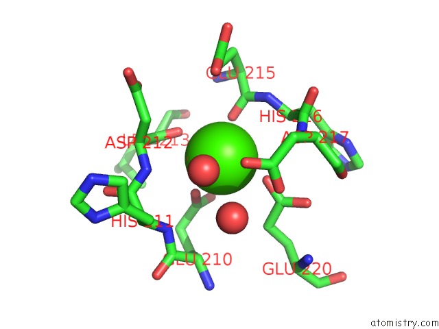
Mono view
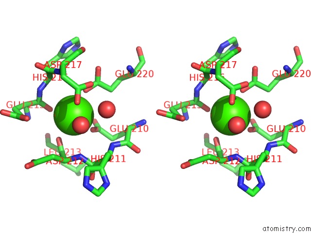
Stereo pair view
Mono view
Stereo pair view
A full contact list of Calcium with other atoms in the Ca binding
site number 1 of Crystal Structure of Active Site Inhibited Coagulation Factor Viia in Complex with Soluble Tissue Factor within 5.0Å range:
|
Reference:
K.S.Larsen,
H.Ostergaard,
J.R.Bjelke,
O.H.Olsen,
H.B.Rasmussen,
L.Christensen,
B.B.Kragelund,
H.R.Stennicke.
Engineering the Substrate and Inhibitor Specificities of Human Coagulation Factor Viia Biochem.J. V. 405 429 2007.
ISSN: ISSN 0264-6021
PubMed: 17456045
DOI: 10.1042/BJ20061901
Page generated: Tue Jul 8 07:47:09 2025
ISSN: ISSN 0264-6021
PubMed: 17456045
DOI: 10.1042/BJ20061901
Last articles
Gd in 4AHWGd in 4AHZ
Gd in 4AHX
Gd in 3ZTY
Gd in 4AHY
Gd in 3ZXS
Gd in 3VDZ
Gd in 3VX0
Gd in 3TXM
Gd in 3Q4I