Calcium »
PDB 2psr-2q91 »
2pwb »
Calcium in PDB 2pwb: Crystal Structure of the Complex of Proteinase K with Coumarin at 1.9 A Resolution
Enzymatic activity of Crystal Structure of the Complex of Proteinase K with Coumarin at 1.9 A Resolution
All present enzymatic activity of Crystal Structure of the Complex of Proteinase K with Coumarin at 1.9 A Resolution:
3.4.21.64;
3.4.21.64;
Protein crystallography data
The structure of Crystal Structure of the Complex of Proteinase K with Coumarin at 1.9 A Resolution, PDB code: 2pwb
was solved by
A.K.Singh,
N.Singh,
M.Sinha,
S.Sharma,
P.Kaur,
T.P.Singh,
with X-Ray Crystallography technique. A brief refinement statistics is given in the table below:
| Resolution Low / High (Å) | 57.74 / 1.90 |
| Space group | P 43 21 2 |
| Cell size a, b, c (Å), α, β, γ (°) | 68.300, 68.300, 108.384, 90.00, 90.00, 90.00 |
| R / Rfree (%) | 14.2 / 17.3 |
Calcium Binding Sites:
The binding sites of Calcium atom in the Crystal Structure of the Complex of Proteinase K with Coumarin at 1.9 A Resolution
(pdb code 2pwb). This binding sites where shown within
5.0 Angstroms radius around Calcium atom.
In total 2 binding sites of Calcium where determined in the Crystal Structure of the Complex of Proteinase K with Coumarin at 1.9 A Resolution, PDB code: 2pwb:
Jump to Calcium binding site number: 1; 2;
In total 2 binding sites of Calcium where determined in the Crystal Structure of the Complex of Proteinase K with Coumarin at 1.9 A Resolution, PDB code: 2pwb:
Jump to Calcium binding site number: 1; 2;
Calcium binding site 1 out of 2 in 2pwb
Go back to
Calcium binding site 1 out
of 2 in the Crystal Structure of the Complex of Proteinase K with Coumarin at 1.9 A Resolution
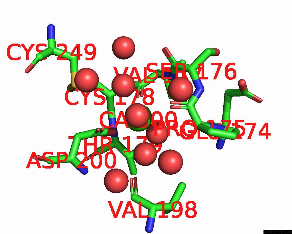
Mono view
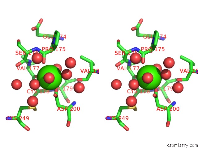
Stereo pair view
Mono view
Stereo pair view
A full contact list of Calcium with other atoms in the Ca binding
site number 1 of Crystal Structure of the Complex of Proteinase K with Coumarin at 1.9 A Resolution within 5.0Å range:
|
Calcium binding site 2 out of 2 in 2pwb
Go back to
Calcium binding site 2 out
of 2 in the Crystal Structure of the Complex of Proteinase K with Coumarin at 1.9 A Resolution
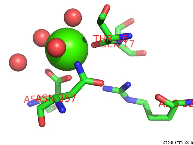
Mono view
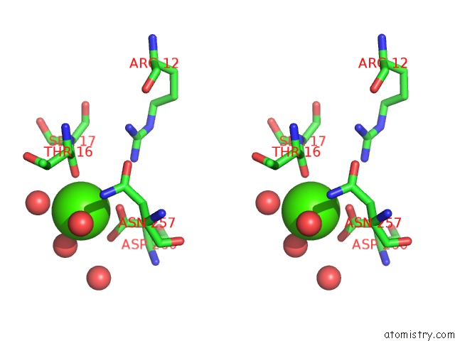
Stereo pair view
Mono view
Stereo pair view
A full contact list of Calcium with other atoms in the Ca binding
site number 2 of Crystal Structure of the Complex of Proteinase K with Coumarin at 1.9 A Resolution within 5.0Å range:
|
Reference:
A.K.Singh,
N.Singh,
M.Sinha,
S.Sharma,
P.Kaur,
T.P.Singh.
Crystal Structure of the Complex of Proteinase K with Coumarin at 1.9A Resolution To Be Published.
Page generated: Tue Jul 8 07:47:50 2025
Last articles
Gd in 5OERGd in 5TC9
Gd in 5N35
Gd in 4PHB
Gd in 5FIS
Gd in 5IWT
Gd in 5FBH
Gd in 4X8I
Gd in 5F6T
Gd in 5C6L