Calcium »
PDB 2q97-2qwc »
2qng »
Calcium in PDB 2qng: Crystal Structure of Unknown Function Protein SAV2460
Protein crystallography data
The structure of Crystal Structure of Unknown Function Protein SAV2460, PDB code: 2qng
was solved by
C.Chang,
X.Xu,
H.Zheng,
A.Savchenko,
A.M.Edwards,
A.Joachimiak,
Midwestcenter For Structural Genomics (Mcsg),
with X-Ray Crystallography technique. A brief refinement statistics is given in the table below:
| Resolution Low / High (Å) | 49.09 / 1.40 |
| Space group | P 1 21 1 |
| Cell size a, b, c (Å), α, β, γ (°) | 43.231, 40.701, 49.436, 90.00, 96.66, 90.00 |
| R / Rfree (%) | 16.5 / 18.6 |
Calcium Binding Sites:
The binding sites of Calcium atom in the Crystal Structure of Unknown Function Protein SAV2460
(pdb code 2qng). This binding sites where shown within
5.0 Angstroms radius around Calcium atom.
In total 2 binding sites of Calcium where determined in the Crystal Structure of Unknown Function Protein SAV2460, PDB code: 2qng:
Jump to Calcium binding site number: 1; 2;
In total 2 binding sites of Calcium where determined in the Crystal Structure of Unknown Function Protein SAV2460, PDB code: 2qng:
Jump to Calcium binding site number: 1; 2;
Calcium binding site 1 out of 2 in 2qng
Go back to
Calcium binding site 1 out
of 2 in the Crystal Structure of Unknown Function Protein SAV2460
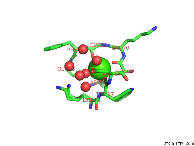
Mono view
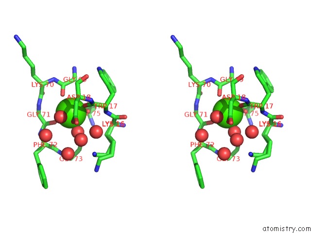
Stereo pair view
Mono view
Stereo pair view
A full contact list of Calcium with other atoms in the Ca binding
site number 1 of Crystal Structure of Unknown Function Protein SAV2460 within 5.0Å range:
|
Calcium binding site 2 out of 2 in 2qng
Go back to
Calcium binding site 2 out
of 2 in the Crystal Structure of Unknown Function Protein SAV2460
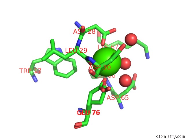
Mono view
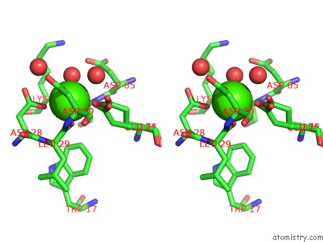
Stereo pair view
Mono view
Stereo pair view
A full contact list of Calcium with other atoms in the Ca binding
site number 2 of Crystal Structure of Unknown Function Protein SAV2460 within 5.0Å range:
|
Reference:
C.Chang,
X.Xu,
H.Zheng,
A.Savchenko,
A.M.Edwards,
A.Joachimiak.
Crystal Structure of SAV2460. To Be Published.
Page generated: Tue Jul 8 07:54:41 2025
Last articles
Mg in 5JI2Mg in 5JIC
Mg in 5JDA
Mg in 5JCO
Mg in 5JH7
Mg in 5JD9
Mg in 5JFC
Mg in 5JCB
Mg in 5JCZ
Mg in 5JCP