Calcium »
PDB 2qwd-2rex »
2r1d »
Calcium in PDB 2r1d: Crystal Structure of Rat Neurexin 1BETA in the CA2+ Containing Form
Protein crystallography data
The structure of Crystal Structure of Rat Neurexin 1BETA in the CA2+ Containing Form, PDB code: 2r1d
was solved by
G.Rudenko,
with X-Ray Crystallography technique. A brief refinement statistics is given in the table below:
| Resolution Low / High (Å) | 20.00 / 2.60 |
| Space group | P 21 21 2 |
| Cell size a, b, c (Å), α, β, γ (°) | 116.683, 195.719, 103.867, 90.00, 90.00, 90.00 |
| R / Rfree (%) | 20.3 / 24.4 |
Calcium Binding Sites:
The binding sites of Calcium atom in the Crystal Structure of Rat Neurexin 1BETA in the CA2+ Containing Form
(pdb code 2r1d). This binding sites where shown within
5.0 Angstroms radius around Calcium atom.
In total 3 binding sites of Calcium where determined in the Crystal Structure of Rat Neurexin 1BETA in the CA2+ Containing Form, PDB code: 2r1d:
Jump to Calcium binding site number: 1; 2; 3;
In total 3 binding sites of Calcium where determined in the Crystal Structure of Rat Neurexin 1BETA in the CA2+ Containing Form, PDB code: 2r1d:
Jump to Calcium binding site number: 1; 2; 3;
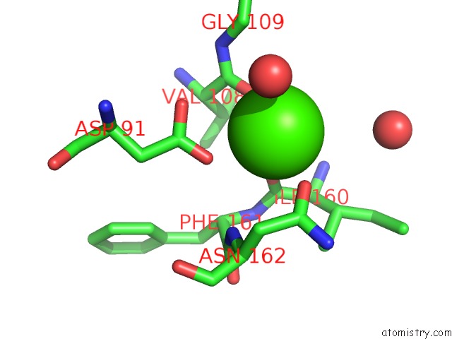
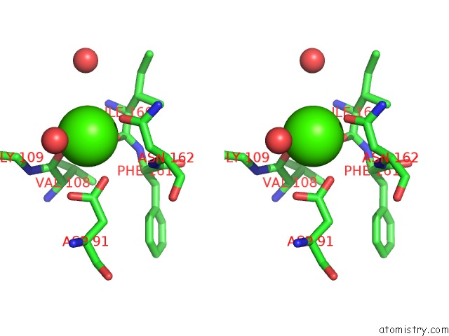
Calcium binding site 1 out of 3 in 2r1d
Go back to
Calcium binding site 1 out
of 3 in the Crystal Structure of Rat Neurexin 1BETA in the CA2+ Containing Form
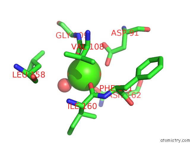
Mono view
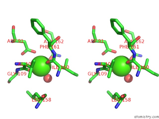
Stereo pair view
Mono view
Stereo pair view
A full contact list of Calcium with other atoms in the Ca binding
site number 1 of Crystal Structure of Rat Neurexin 1BETA in the CA2+ Containing Form within 5.0Å range:
|
Calcium binding site 2 out of 3 in 2r1d
Go back to
Calcium binding site 2 out
of 3 in the Crystal Structure of Rat Neurexin 1BETA in the CA2+ Containing Form
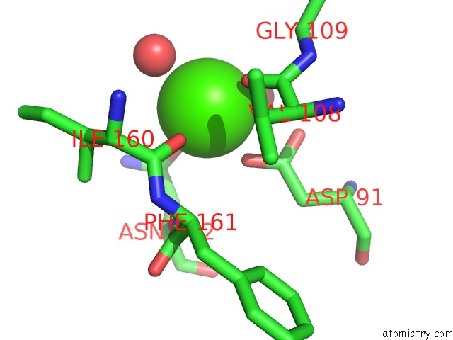
Mono view
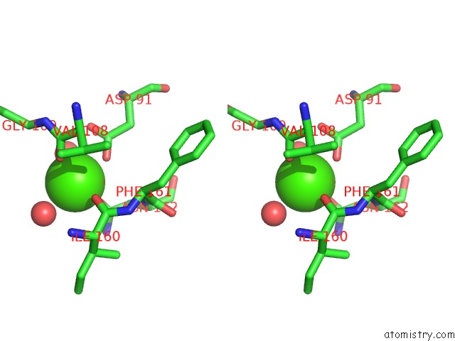
Stereo pair view
Mono view
Stereo pair view
A full contact list of Calcium with other atoms in the Ca binding
site number 2 of Crystal Structure of Rat Neurexin 1BETA in the CA2+ Containing Form within 5.0Å range:
|
Calcium binding site 3 out of 3 in 2r1d
Go back to
Calcium binding site 3 out
of 3 in the Crystal Structure of Rat Neurexin 1BETA in the CA2+ Containing Form
Mono view
Stereo pair view
Mono view
Stereo pair view
A full contact list of Calcium with other atoms in the Ca binding
site number 3 of Crystal Structure of Rat Neurexin 1BETA in the CA2+ Containing Form within 5.0Å range:
|
Reference:
K.C.Shen,
D.A.Kuczynska,
I.J.Wu,
B.H.Murray,
L.R.Sheckler,
G.Rudenko.
Regulation of Neurexin 1BETA Tertiary Structure and Ligand Binding Through Alternative Splicing Structure V. 16 422 2008.
ISSN: ISSN 0969-2126
PubMed: 18334217
DOI: 10.1016/J.STR.2008.01.005
Page generated: Tue Jul 8 08:05:13 2025
ISSN: ISSN 0969-2126
PubMed: 18334217
DOI: 10.1016/J.STR.2008.01.005
Last articles
Mg in 4DUYMg in 4DR7
Mg in 4DR6
Mg in 4DR5
Mg in 4DUX
Mg in 4DUW
Mg in 4DUV
Mg in 4DUO
Mg in 4DUG
Mg in 4DTY