Calcium »
PDB 2qwd-2rex »
2r2i »
Calcium in PDB 2r2i: Myristoylated Guanylate Cyclase Activating Protein-1 with Calcium Bound
Protein crystallography data
The structure of Myristoylated Guanylate Cyclase Activating Protein-1 with Calcium Bound, PDB code: 2r2i
was solved by
R.Stephen,
with X-Ray Crystallography technique. A brief refinement statistics is given in the table below:
| Resolution Low / High (Å) | 29.93 / 2.00 |
| Space group | P 41 21 2 |
| Cell size a, b, c (Å), α, β, γ (°) | 73.333, 73.333, 73.229, 90.00, 90.00, 90.00 |
| R / Rfree (%) | 21.8 / 25.1 |
Calcium Binding Sites:
The binding sites of Calcium atom in the Myristoylated Guanylate Cyclase Activating Protein-1 with Calcium Bound
(pdb code 2r2i). This binding sites where shown within
5.0 Angstroms radius around Calcium atom.
In total 3 binding sites of Calcium where determined in the Myristoylated Guanylate Cyclase Activating Protein-1 with Calcium Bound, PDB code: 2r2i:
Jump to Calcium binding site number: 1; 2; 3;
In total 3 binding sites of Calcium where determined in the Myristoylated Guanylate Cyclase Activating Protein-1 with Calcium Bound, PDB code: 2r2i:
Jump to Calcium binding site number: 1; 2; 3;
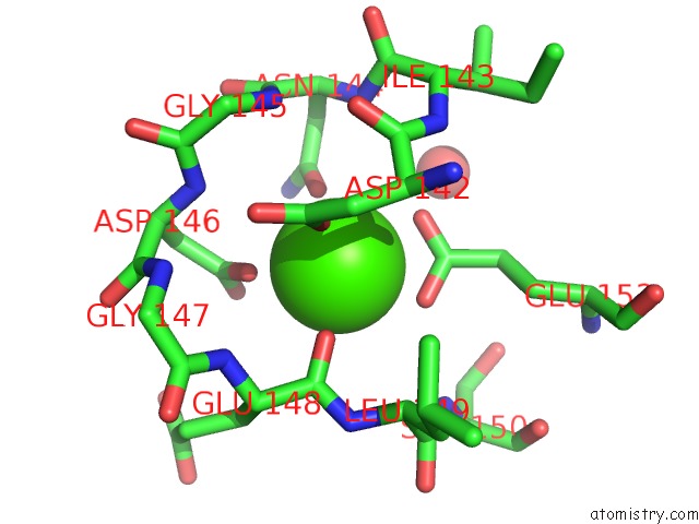
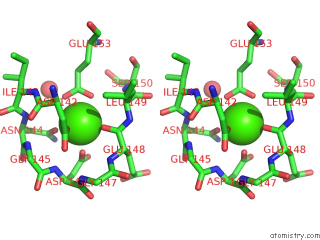
Calcium binding site 1 out of 3 in 2r2i
Go back to
Calcium binding site 1 out
of 3 in the Myristoylated Guanylate Cyclase Activating Protein-1 with Calcium Bound
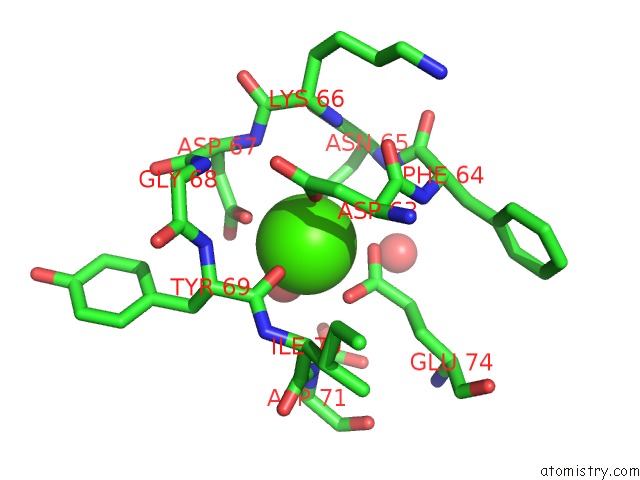
Mono view
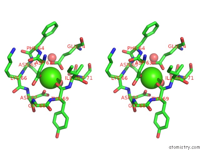
Stereo pair view
Mono view
Stereo pair view
A full contact list of Calcium with other atoms in the Ca binding
site number 1 of Myristoylated Guanylate Cyclase Activating Protein-1 with Calcium Bound within 5.0Å range:
|
Calcium binding site 2 out of 3 in 2r2i
Go back to
Calcium binding site 2 out
of 3 in the Myristoylated Guanylate Cyclase Activating Protein-1 with Calcium Bound
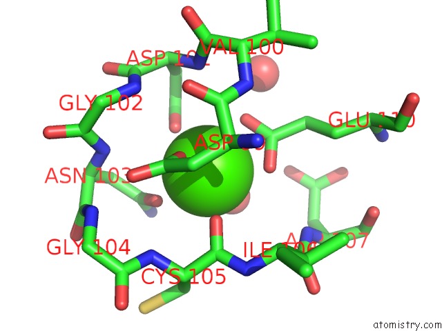
Mono view
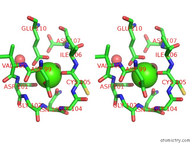
Stereo pair view
Mono view
Stereo pair view
A full contact list of Calcium with other atoms in the Ca binding
site number 2 of Myristoylated Guanylate Cyclase Activating Protein-1 with Calcium Bound within 5.0Å range:
|
Calcium binding site 3 out of 3 in 2r2i
Go back to
Calcium binding site 3 out
of 3 in the Myristoylated Guanylate Cyclase Activating Protein-1 with Calcium Bound
Mono view
Stereo pair view
Mono view
Stereo pair view
A full contact list of Calcium with other atoms in the Ca binding
site number 3 of Myristoylated Guanylate Cyclase Activating Protein-1 with Calcium Bound within 5.0Å range:
|
Reference:
R.Stephen,
G.Bereta,
M.Golczak,
K.Palczewski,
M.C.Sousa.
Stabilizing Function For Myristoyl Group Revealed By the Crystal Structure of A Neuronal Calcium Sensor, Guanylate Cyclase-Activating Protein 1. Structure V. 15 1392 2007.
ISSN: ISSN 0969-2126
PubMed: 17997965
DOI: 10.1016/J.STR.2007.09.013
Page generated: Fri Jul 12 15:44:54 2024
ISSN: ISSN 0969-2126
PubMed: 17997965
DOI: 10.1016/J.STR.2007.09.013
Last articles
Zn in 9MJ5Zn in 9HNW
Zn in 9G0L
Zn in 9FNE
Zn in 9DZN
Zn in 9E0I
Zn in 9D32
Zn in 9DAK
Zn in 8ZXC
Zn in 8ZUF