Calcium »
PDB 2qwd-2rex »
2re1 »
Calcium in PDB 2re1: Crystal Structure of Aspartokinase Alpha and Beta Subunits
Enzymatic activity of Crystal Structure of Aspartokinase Alpha and Beta Subunits
All present enzymatic activity of Crystal Structure of Aspartokinase Alpha and Beta Subunits:
2.7.2.4;
2.7.2.4;
Protein crystallography data
The structure of Crystal Structure of Aspartokinase Alpha and Beta Subunits, PDB code: 2re1
was solved by
C.Chang,
H.Li,
M.Gu,
A.Joachimiak,
Midwest Center For Structural Genomics(Mcsg),
with X-Ray Crystallography technique. A brief refinement statistics is given in the table below:
| Resolution Low / High (Å) | 44.46 / 2.75 |
| Space group | P 41 21 2 |
| Cell size a, b, c (Å), α, β, γ (°) | 61.806, 61.806, 191.901, 90.00, 90.00, 90.00 |
| R / Rfree (%) | 23.4 / 27.6 |
Calcium Binding Sites:
The binding sites of Calcium atom in the Crystal Structure of Aspartokinase Alpha and Beta Subunits
(pdb code 2re1). This binding sites where shown within
5.0 Angstroms radius around Calcium atom.
In total 2 binding sites of Calcium where determined in the Crystal Structure of Aspartokinase Alpha and Beta Subunits, PDB code: 2re1:
Jump to Calcium binding site number: 1; 2;
In total 2 binding sites of Calcium where determined in the Crystal Structure of Aspartokinase Alpha and Beta Subunits, PDB code: 2re1:
Jump to Calcium binding site number: 1; 2;
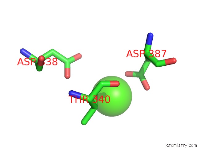
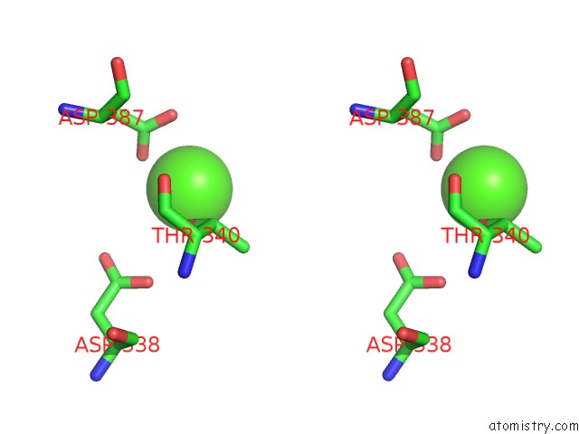
Calcium binding site 1 out of 2 in 2re1
Go back to
Calcium binding site 1 out
of 2 in the Crystal Structure of Aspartokinase Alpha and Beta Subunits

Mono view

Stereo pair view
Mono view
Stereo pair view
A full contact list of Calcium with other atoms in the Ca binding
site number 1 of Crystal Structure of Aspartokinase Alpha and Beta Subunits within 5.0Å range:
|
Calcium binding site 2 out of 2 in 2re1
Go back to
Calcium binding site 2 out
of 2 in the Crystal Structure of Aspartokinase Alpha and Beta Subunits
Mono view
Stereo pair view
Mono view
Stereo pair view
A full contact list of Calcium with other atoms in the Ca binding
site number 2 of Crystal Structure of Aspartokinase Alpha and Beta Subunits within 5.0Å range:
|
Reference:
C.Chang,
H.Li,
M.Gu,
A.Joachimiak.
Crystal Structure of Aspartokinase Alpha and Beta Subunits. To Be Published.
Page generated: Tue Jul 8 08:11:59 2025
Last articles
Mg in 5SB6Mg in 5SB4
Mg in 5SB5
Mg in 5SB3
Mg in 5SA3
Mg in 5SA2
Mg in 5SA1
Mg in 5SA0
Mg in 5S9Y
Mg in 5S9Z