Calcium »
PDB 2qwd-2rex »
2rex »
Calcium in PDB 2rex: Crystal Structure of the Effector Domain of PLXNB1 Bound with RND1 Gtpase
Protein crystallography data
The structure of Crystal Structure of the Effector Domain of PLXNB1 Bound with RND1 Gtpase, PDB code: 2rex
was solved by
Y.Tong,
W.Tempel,
L.Shen,
C.H.Arrowsmith,
A.M.Edwards,
M.Sundstrom,
J.Weigelt,
A.Bochkarev,
H.Park,
Structural Genomics Consortium (Sgc),
with X-Ray Crystallography technique. A brief refinement statistics is given in the table below:
| Resolution Low / High (Å) | 20.00 / 2.30 |
| Space group | C 1 2 1 |
| Cell size a, b, c (Å), α, β, γ (°) | 150.136, 71.653, 101.885, 90.00, 128.36, 90.00 |
| R / Rfree (%) | 21.5 / 24.8 |
Other elements in 2rex:
The structure of Crystal Structure of the Effector Domain of PLXNB1 Bound with RND1 Gtpase also contains other interesting chemical elements:
| Magnesium | (Mg) | 2 atoms |
Calcium Binding Sites:
The binding sites of Calcium atom in the Crystal Structure of the Effector Domain of PLXNB1 Bound with RND1 Gtpase
(pdb code 2rex). This binding sites where shown within
5.0 Angstroms radius around Calcium atom.
In total 2 binding sites of Calcium where determined in the Crystal Structure of the Effector Domain of PLXNB1 Bound with RND1 Gtpase, PDB code: 2rex:
Jump to Calcium binding site number: 1; 2;
In total 2 binding sites of Calcium where determined in the Crystal Structure of the Effector Domain of PLXNB1 Bound with RND1 Gtpase, PDB code: 2rex:
Jump to Calcium binding site number: 1; 2;
Calcium binding site 1 out of 2 in 2rex
Go back to
Calcium binding site 1 out
of 2 in the Crystal Structure of the Effector Domain of PLXNB1 Bound with RND1 Gtpase
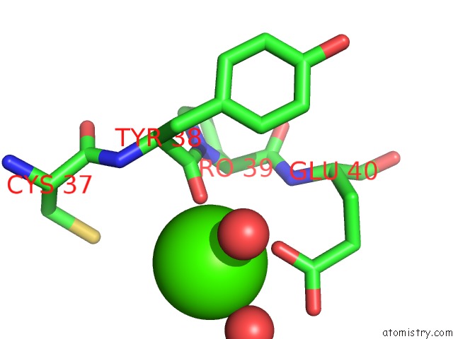
Mono view
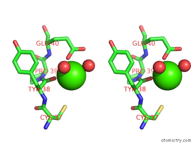
Stereo pair view
Mono view
Stereo pair view
A full contact list of Calcium with other atoms in the Ca binding
site number 1 of Crystal Structure of the Effector Domain of PLXNB1 Bound with RND1 Gtpase within 5.0Å range:
|
Calcium binding site 2 out of 2 in 2rex
Go back to
Calcium binding site 2 out
of 2 in the Crystal Structure of the Effector Domain of PLXNB1 Bound with RND1 Gtpase
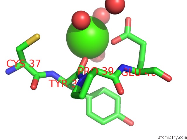
Mono view
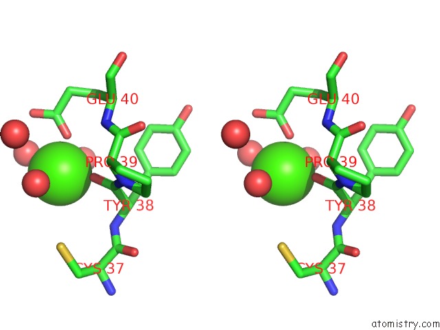
Stereo pair view
Mono view
Stereo pair view
A full contact list of Calcium with other atoms in the Ca binding
site number 2 of Crystal Structure of the Effector Domain of PLXNB1 Bound with RND1 Gtpase within 5.0Å range:
|
Reference:
Y.Tong,
W.Tempel,
L.Shen,
C.H.Arrowsmith,
A.M.Edwards,
M.Sundstrom,
J.Weigelt,
A.Bochkarev,
H.Park.
Crystal Structure of the Effector Domain of PLXNB1 Bound with RND1 Gtpase. To Be Published.
Page generated: Tue Jul 8 08:12:03 2025
Last articles
Mg in 2WJHMg in 2WJI
Mg in 2WIC
Mg in 2WIA
Mg in 2WI1
Mg in 2WI3
Mg in 2WGH
Mg in 2WFI
Mg in 2WHE
Mg in 2WFJ