Calcium »
PDB 2tga-2v5c »
2uvw »
Calcium in PDB 2uvw: Crystal Structures of Mutant DPO4 Dna Polymerases with 8- Oxog Containing Dna Template-Primer Constructs
Enzymatic activity of Crystal Structures of Mutant DPO4 Dna Polymerases with 8- Oxog Containing Dna Template-Primer Constructs
All present enzymatic activity of Crystal Structures of Mutant DPO4 Dna Polymerases with 8- Oxog Containing Dna Template-Primer Constructs:
2.7.7.7;
2.7.7.7;
Protein crystallography data
The structure of Crystal Structures of Mutant DPO4 Dna Polymerases with 8- Oxog Containing Dna Template-Primer Constructs, PDB code: 2uvw
was solved by
A.Irimia,
M.Egli,
with X-Ray Crystallography technique. A brief refinement statistics is given in the table below:
| Resolution Low / High (Å) | 30.81 / 2.09 |
| Space group | P 21 21 2 |
| Cell size a, b, c (Å), α, β, γ (°) | 96.778, 103.728, 52.915, 90.00, 90.00, 90.00 |
| R / Rfree (%) | 24.9 / 27.4 |
Calcium Binding Sites:
The binding sites of Calcium atom in the Crystal Structures of Mutant DPO4 Dna Polymerases with 8- Oxog Containing Dna Template-Primer Constructs
(pdb code 2uvw). This binding sites where shown within
5.0 Angstroms radius around Calcium atom.
In total 3 binding sites of Calcium where determined in the Crystal Structures of Mutant DPO4 Dna Polymerases with 8- Oxog Containing Dna Template-Primer Constructs, PDB code: 2uvw:
Jump to Calcium binding site number: 1; 2; 3;
In total 3 binding sites of Calcium where determined in the Crystal Structures of Mutant DPO4 Dna Polymerases with 8- Oxog Containing Dna Template-Primer Constructs, PDB code: 2uvw:
Jump to Calcium binding site number: 1; 2; 3;
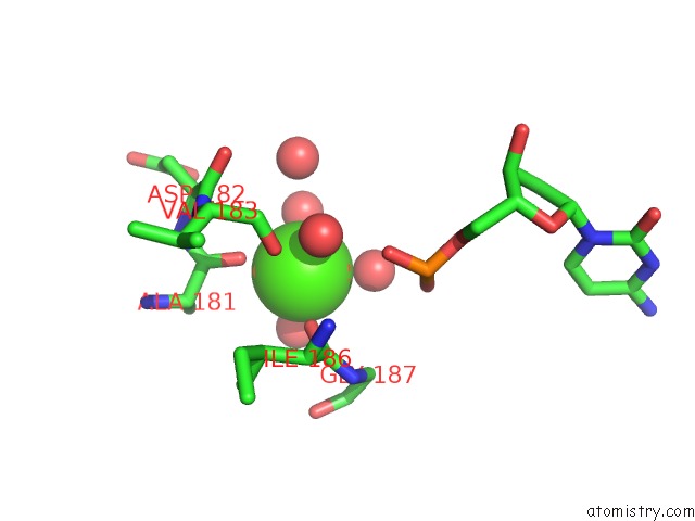
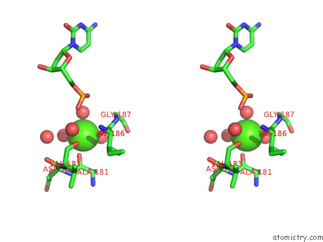
Calcium binding site 1 out of 3 in 2uvw
Go back to
Calcium binding site 1 out
of 3 in the Crystal Structures of Mutant DPO4 Dna Polymerases with 8- Oxog Containing Dna Template-Primer Constructs
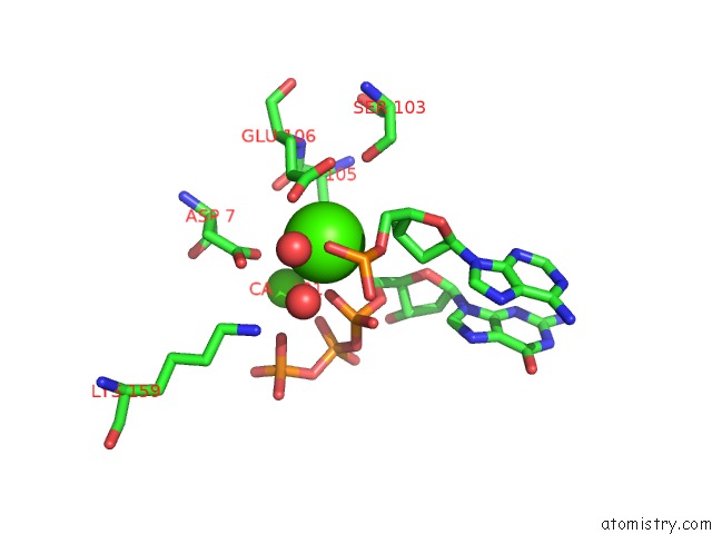
Mono view
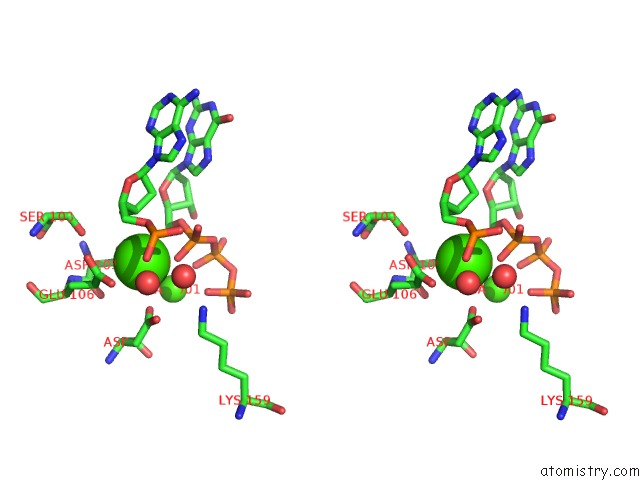
Stereo pair view
Mono view
Stereo pair view
A full contact list of Calcium with other atoms in the Ca binding
site number 1 of Crystal Structures of Mutant DPO4 Dna Polymerases with 8- Oxog Containing Dna Template-Primer Constructs within 5.0Å range:
|
Calcium binding site 2 out of 3 in 2uvw
Go back to
Calcium binding site 2 out
of 3 in the Crystal Structures of Mutant DPO4 Dna Polymerases with 8- Oxog Containing Dna Template-Primer Constructs
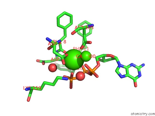
Mono view
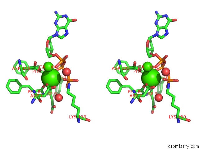
Stereo pair view
Mono view
Stereo pair view
A full contact list of Calcium with other atoms in the Ca binding
site number 2 of Crystal Structures of Mutant DPO4 Dna Polymerases with 8- Oxog Containing Dna Template-Primer Constructs within 5.0Å range:
|
Calcium binding site 3 out of 3 in 2uvw
Go back to
Calcium binding site 3 out
of 3 in the Crystal Structures of Mutant DPO4 Dna Polymerases with 8- Oxog Containing Dna Template-Primer Constructs
Mono view
Stereo pair view
Mono view
Stereo pair view
A full contact list of Calcium with other atoms in the Ca binding
site number 3 of Crystal Structures of Mutant DPO4 Dna Polymerases with 8- Oxog Containing Dna Template-Primer Constructs within 5.0Å range:
|
Reference:
R.L.Eoff,
A.Irimia,
K.C.Angel,
M.Egli,
F.P.Guengerich.
Hydrogen Bonding of 7,8-Dihydro-8- Oxodeoxyguanosine with A Charged Residue in the Little Finger Domain Determines Miscoding Events in Sulfolobus Solfataricus Dna Polymerase DPO4. J.Biol.Chem. V. 282 19831 2007.
ISSN: ISSN 0021-9258
PubMed: 17468100
DOI: 10.1074/JBC.M702290200
Page generated: Tue Jul 8 08:27:19 2025
ISSN: ISSN 0021-9258
PubMed: 17468100
DOI: 10.1074/JBC.M702290200
Last articles
K in 8S1UK in 8SDA
K in 8SFZ
K in 8SD3
K in 8S3E
K in 8S4S
K in 8S1X
K in 8RUH
K in 8RUJ
K in 8RUI