Calcium »
PDB 2tga-2v5c »
2ux1 »
Calcium in PDB 2ux1: Identification of Two Zinc-Binding Sites in the Streptococcus Suis Dpr Protein
Protein crystallography data
The structure of Identification of Two Zinc-Binding Sites in the Streptococcus Suis Dpr Protein, PDB code: 2ux1
was solved by
H.Havukainen,
A.Kauko,
A.T.Pulliainen,
S.Haataja,
W.Meyer-Klaucke,
J.Finne,
A.C.Papageorgiou,
with X-Ray Crystallography technique. A brief refinement statistics is given in the table below:
| Resolution Low / High (Å) | 19.97 / 1.80 |
| Space group | P 21 21 21 |
| Cell size a, b, c (Å), α, β, γ (°) | 104.520, 137.690, 142.060, 90.00, 90.00, 90.00 |
| R / Rfree (%) | 20.6 / 24.8 |
Other elements in 2ux1:
The structure of Identification of Two Zinc-Binding Sites in the Streptococcus Suis Dpr Protein also contains other interesting chemical elements:
| Chlorine | (Cl) | 2 atoms |
| Zinc | (Zn) | 24 atoms |
Calcium Binding Sites:
The binding sites of Calcium atom in the Identification of Two Zinc-Binding Sites in the Streptococcus Suis Dpr Protein
(pdb code 2ux1). This binding sites where shown within
5.0 Angstroms radius around Calcium atom.
In total 4 binding sites of Calcium where determined in the Identification of Two Zinc-Binding Sites in the Streptococcus Suis Dpr Protein, PDB code: 2ux1:
Jump to Calcium binding site number: 1; 2; 3; 4;
In total 4 binding sites of Calcium where determined in the Identification of Two Zinc-Binding Sites in the Streptococcus Suis Dpr Protein, PDB code: 2ux1:
Jump to Calcium binding site number: 1; 2; 3; 4;
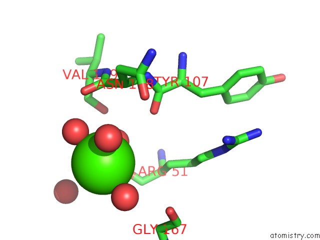
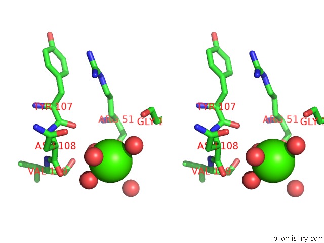
Calcium binding site 1 out of 4 in 2ux1
Go back to
Calcium binding site 1 out
of 4 in the Identification of Two Zinc-Binding Sites in the Streptococcus Suis Dpr Protein

Mono view

Stereo pair view
Mono view
Stereo pair view
A full contact list of Calcium with other atoms in the Ca binding
site number 1 of Identification of Two Zinc-Binding Sites in the Streptococcus Suis Dpr Protein within 5.0Å range:
|
Calcium binding site 2 out of 4 in 2ux1
Go back to
Calcium binding site 2 out
of 4 in the Identification of Two Zinc-Binding Sites in the Streptococcus Suis Dpr Protein

Mono view

Stereo pair view
Mono view
Stereo pair view
A full contact list of Calcium with other atoms in the Ca binding
site number 2 of Identification of Two Zinc-Binding Sites in the Streptococcus Suis Dpr Protein within 5.0Å range:
|
Calcium binding site 3 out of 4 in 2ux1
Go back to
Calcium binding site 3 out
of 4 in the Identification of Two Zinc-Binding Sites in the Streptococcus Suis Dpr Protein
Mono view
Stereo pair view
Mono view
Stereo pair view
A full contact list of Calcium with other atoms in the Ca binding
site number 3 of Identification of Two Zinc-Binding Sites in the Streptococcus Suis Dpr Protein within 5.0Å range:
|
Calcium binding site 4 out of 4 in 2ux1
Go back to
Calcium binding site 4 out
of 4 in the Identification of Two Zinc-Binding Sites in the Streptococcus Suis Dpr Protein

Mono view

Stereo pair view
Mono view
Stereo pair view
A full contact list of Calcium with other atoms in the Ca binding
site number 4 of Identification of Two Zinc-Binding Sites in the Streptococcus Suis Dpr Protein within 5.0Å range:
|
Reference:
H.Havukainen,
S.Haataja,
A.Kauko,
A.T.Pulliainen,
A.Salminen,
T.Haikarainen,
J.Finne,
A.C.Papageorgiou.
Structural Basis of the Zinc- and Terbium-Mediated Inhibition of Ferroxidase Activity in Dps Ferritin- Like Proteins. Protein Sci. V. 17 1513 2008.
ISSN: ISSN 0961-8368
PubMed: 18552126
DOI: 10.1110/PS.036236.108
Page generated: Tue Jul 8 08:27:40 2025
ISSN: ISSN 0961-8368
PubMed: 18552126
DOI: 10.1110/PS.036236.108
Last articles
K in 5K0NK in 5K6R
K in 5K0L
K in 5JZX
K in 5K01
K in 5K03
K in 5J9P
K in 5JPR
K in 5JQR
K in 5JGZ