Calcium »
PDB 2tga-2v5c »
2v51 »
Calcium in PDB 2v51: Structure of Mal-RPEL1 Complexed to Actin
Protein crystallography data
The structure of Structure of Mal-RPEL1 Complexed to Actin, PDB code: 2v51
was solved by
S.Mouilleron,
S.Guettler,
C.A.Langer,
R.Treisman,
N.Q.Mcdonald,
with X-Ray Crystallography technique. A brief refinement statistics is given in the table below:
| Resolution Low / High (Å) | 20.00 / 2.35 |
| Space group | P 1 21 1 |
| Cell size a, b, c (Å), α, β, γ (°) | 53.092, 59.071, 166.203, 90.00, 90.06, 90.00 |
| R / Rfree (%) | 18.3 / 24.8 |
Calcium Binding Sites:
The binding sites of Calcium atom in the Structure of Mal-RPEL1 Complexed to Actin
(pdb code 2v51). This binding sites where shown within
5.0 Angstroms radius around Calcium atom.
In total 2 binding sites of Calcium where determined in the Structure of Mal-RPEL1 Complexed to Actin, PDB code: 2v51:
Jump to Calcium binding site number: 1; 2;
In total 2 binding sites of Calcium where determined in the Structure of Mal-RPEL1 Complexed to Actin, PDB code: 2v51:
Jump to Calcium binding site number: 1; 2;
Calcium binding site 1 out of 2 in 2v51
Go back to
Calcium binding site 1 out
of 2 in the Structure of Mal-RPEL1 Complexed to Actin
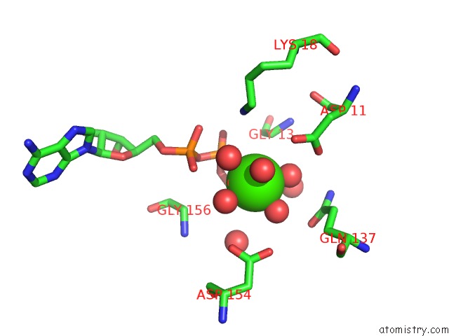
Mono view
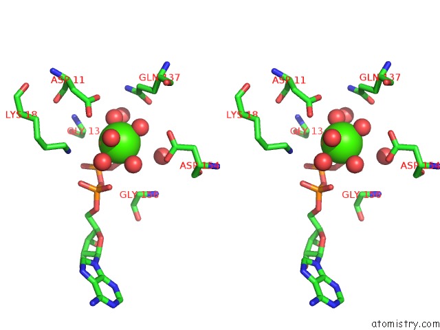
Stereo pair view
Mono view
Stereo pair view
A full contact list of Calcium with other atoms in the Ca binding
site number 1 of Structure of Mal-RPEL1 Complexed to Actin within 5.0Å range:
|
Calcium binding site 2 out of 2 in 2v51
Go back to
Calcium binding site 2 out
of 2 in the Structure of Mal-RPEL1 Complexed to Actin
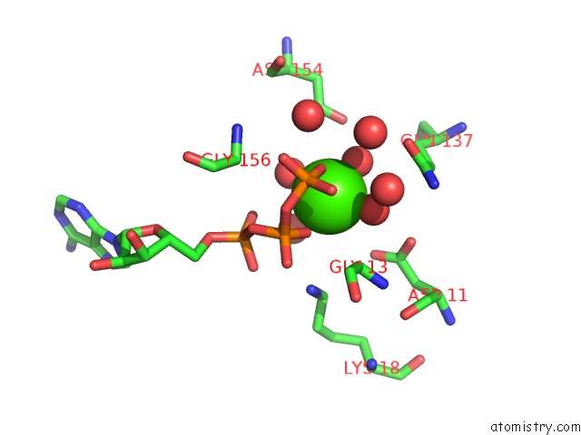
Mono view
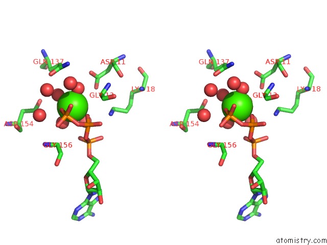
Stereo pair view
Mono view
Stereo pair view
A full contact list of Calcium with other atoms in the Ca binding
site number 2 of Structure of Mal-RPEL1 Complexed to Actin within 5.0Å range:
|
Reference:
S.Mouilleron,
S.Guettler,
C.A.Langer,
R.Treisman,
N.Q.Mcdonald.
Molecular Basis For G-Actin Binding to Rpel Motifs From the Serum Response Factor Coactivator Mal. Embo J. V. 27 3198 2008.
ISSN: ESSN 1460-2075
PubMed: 19008859
DOI: 10.1038/EMBOJ.2008.235
Page generated: Tue Jul 8 08:29:41 2025
ISSN: ESSN 1460-2075
PubMed: 19008859
DOI: 10.1038/EMBOJ.2008.235
Last articles
K in 8YTIK in 8YZ7
K in 8XW8
K in 8XW9
K in 8XTS
K in 8XTR
K in 8XW7
K in 8XW6
K in 8XTQ
K in 8XMI