Calcium »
PDB 2vk5-2vuz »
2vma »
Calcium in PDB 2vma: The Three-Dimensional Structure of the Cytoplasmic Domains of Epsf From the Type 2 Secretion System of Vibrio Cholerae
Protein crystallography data
The structure of The Three-Dimensional Structure of the Cytoplasmic Domains of Epsf From the Type 2 Secretion System of Vibrio Cholerae, PDB code: 2vma
was solved by
J.Abendroth,
K.V.Korotkov,
D.D.Mitchell,
A.Kreger,
W.G.J.Hol,
with X-Ray Crystallography technique. A brief refinement statistics is given in the table below:
| Resolution Low / High (Å) | 46.32 / 1.90 |
| Space group | P 21 21 21 |
| Cell size a, b, c (Å), α, β, γ (°) | 48.816, 54.368, 88.587, 90.00, 90.00, 90.00 |
| R / Rfree (%) | 20.8 / 25.5 |
Other elements in 2vma:
The structure of The Three-Dimensional Structure of the Cytoplasmic Domains of Epsf From the Type 2 Secretion System of Vibrio Cholerae also contains other interesting chemical elements:
| Iodine | (I) | 15 atoms |
Calcium Binding Sites:
The binding sites of Calcium atom in the The Three-Dimensional Structure of the Cytoplasmic Domains of Epsf From the Type 2 Secretion System of Vibrio Cholerae
(pdb code 2vma). This binding sites where shown within
5.0 Angstroms radius around Calcium atom.
In total 3 binding sites of Calcium where determined in the The Three-Dimensional Structure of the Cytoplasmic Domains of Epsf From the Type 2 Secretion System of Vibrio Cholerae, PDB code: 2vma:
Jump to Calcium binding site number: 1; 2; 3;
In total 3 binding sites of Calcium where determined in the The Three-Dimensional Structure of the Cytoplasmic Domains of Epsf From the Type 2 Secretion System of Vibrio Cholerae, PDB code: 2vma:
Jump to Calcium binding site number: 1; 2; 3;
Calcium binding site 1 out of 3 in 2vma
Go back to
Calcium binding site 1 out
of 3 in the The Three-Dimensional Structure of the Cytoplasmic Domains of Epsf From the Type 2 Secretion System of Vibrio Cholerae
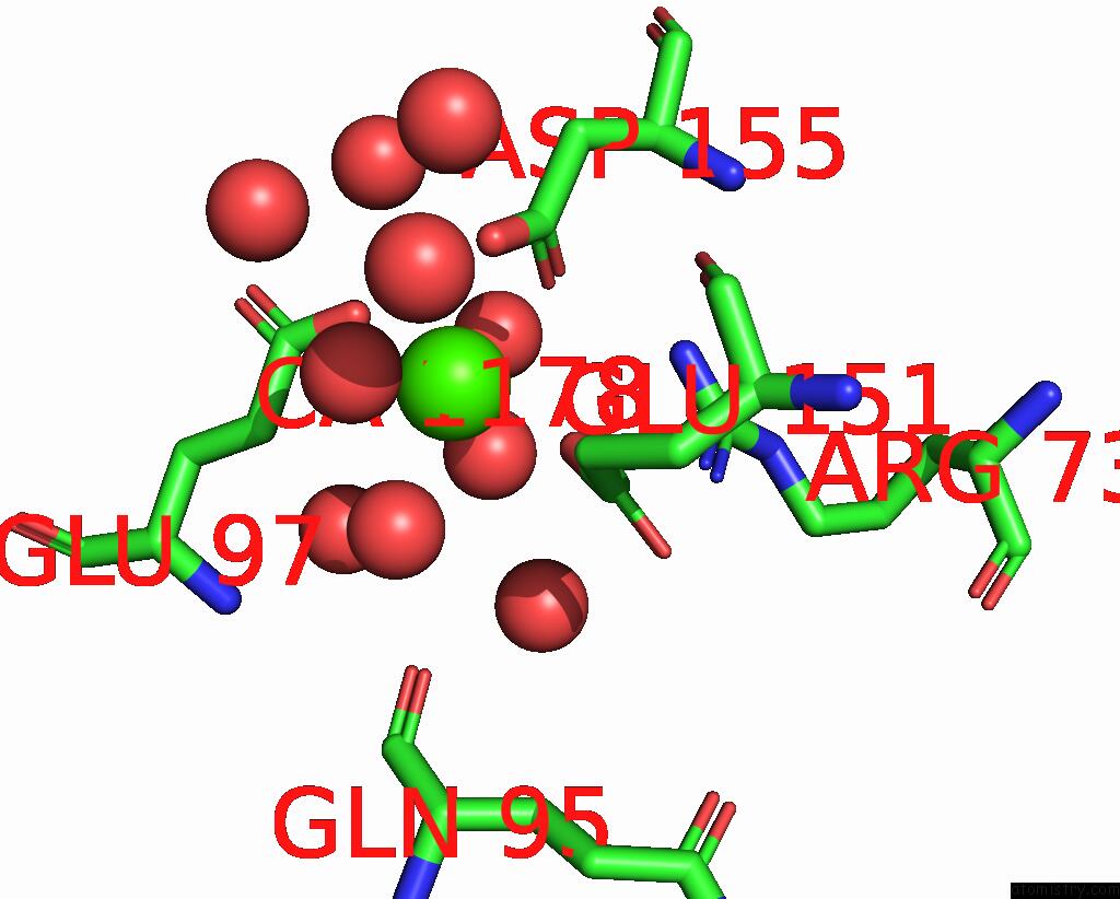
Mono view
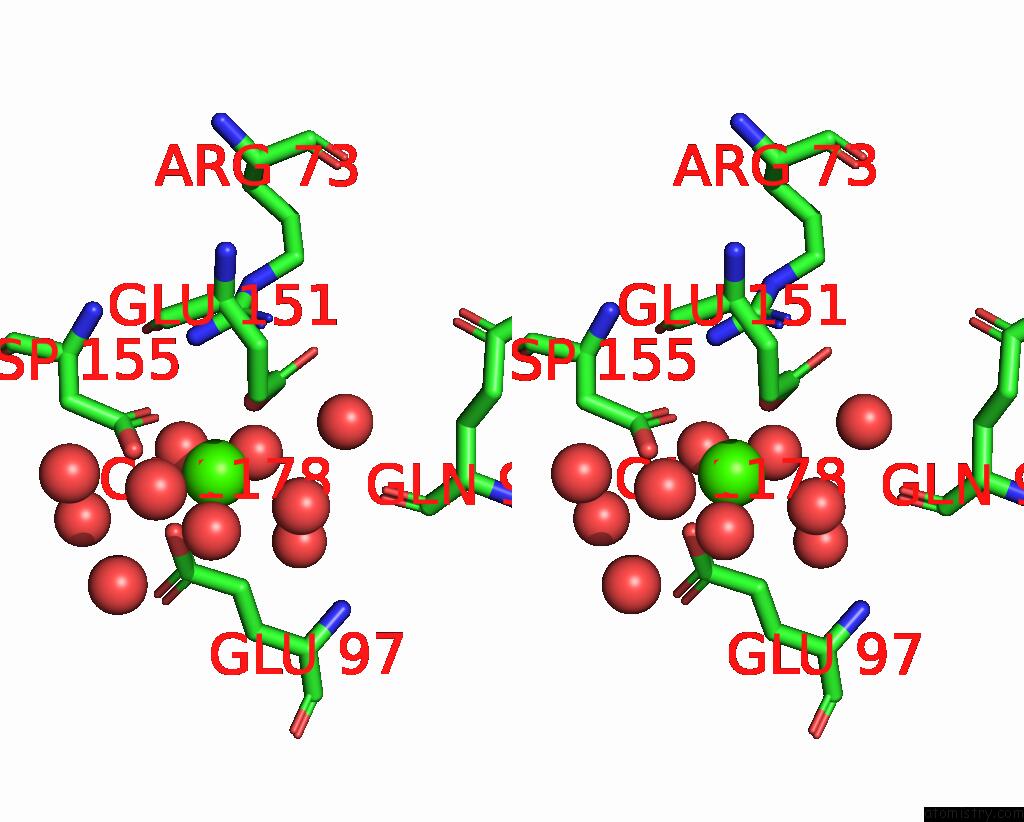
Stereo pair view
Mono view
Stereo pair view
A full contact list of Calcium with other atoms in the Ca binding
site number 1 of The Three-Dimensional Structure of the Cytoplasmic Domains of Epsf From the Type 2 Secretion System of Vibrio Cholerae within 5.0Å range:
|
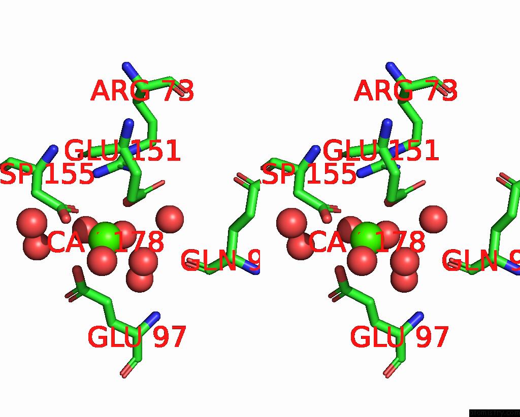
Calcium binding site 2 out of 3 in 2vma
Go back to
Calcium binding site 2 out
of 3 in the The Three-Dimensional Structure of the Cytoplasmic Domains of Epsf From the Type 2 Secretion System of Vibrio Cholerae

Mono view
Stereo pair view
Mono view
Stereo pair view
A full contact list of Calcium with other atoms in the Ca binding
site number 2 of The Three-Dimensional Structure of the Cytoplasmic Domains of Epsf From the Type 2 Secretion System of Vibrio Cholerae within 5.0Å range:
|
Calcium binding site 3 out of 3 in 2vma
Go back to
Calcium binding site 3 out
of 3 in the The Three-Dimensional Structure of the Cytoplasmic Domains of Epsf From the Type 2 Secretion System of Vibrio Cholerae
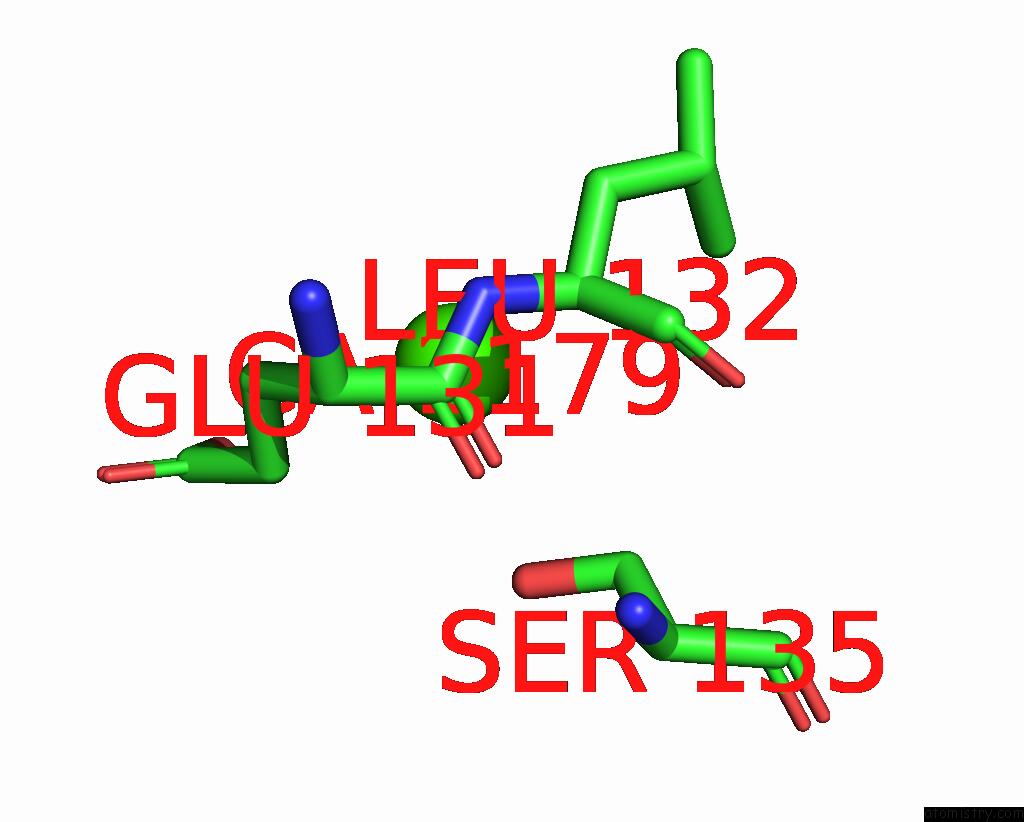
Mono view

Stereo pair view
Mono view
Stereo pair view
A full contact list of Calcium with other atoms in the Ca binding
site number 3 of The Three-Dimensional Structure of the Cytoplasmic Domains of Epsf From the Type 2 Secretion System of Vibrio Cholerae within 5.0Å range:
|
Reference:
J.Abendroth,
D.D.Mitchell,
K.V.Korotkov,
T.L.Johnson,
A.Kreger,
M.Sandkvist,
W.G.Hol.
The Three-Dimensional Structure of the Cytoplasmic Domains of Epsf From the Type 2 Secretion System of Vibrio Cholerae. J.Struct.Biol. V. 166 303 2009.
ISSN: ISSN 1047-8477
PubMed: 19324092
DOI: 10.1016/J.JSB.2009.03.009
Page generated: Tue Jul 8 08:39:55 2025
ISSN: ISSN 1047-8477
PubMed: 19324092
DOI: 10.1016/J.JSB.2009.03.009
Last articles
Na in 4JD5Na in 4JCM
Na in 4JCI
Na in 4JB3
Na in 4JAN
Na in 4J4S
Na in 4J9X
Na in 4J9W
Na in 4J9M
Na in 4J6W