Calcium »
PDB 2vk5-2vuz »
2vmg »
Calcium in PDB 2vmg: The Structure of CBM51 From Clostridium Perfringens GH95 in Complex with Methyl-Galactose
Protein crystallography data
The structure of The Structure of CBM51 From Clostridium Perfringens GH95 in Complex with Methyl-Galactose, PDB code: 2vmg
was solved by
K.Gregg,
R.Finn,
D.W.Abbott,
A.B.Boraston,
with X-Ray Crystallography technique. A brief refinement statistics is given in the table below:
| Resolution Low / High (Å) | 20.00 / 1.90 |
| Space group | P 1 21 1 |
| Cell size a, b, c (Å), α, β, γ (°) | 37.475, 47.948, 38.704, 90.00, 92.00, 90.00 |
| R / Rfree (%) | 23 / 29.6 |
Calcium Binding Sites:
The binding sites of Calcium atom in the The Structure of CBM51 From Clostridium Perfringens GH95 in Complex with Methyl-Galactose
(pdb code 2vmg). This binding sites where shown within
5.0 Angstroms radius around Calcium atom.
In total only one binding site of Calcium was determined in the The Structure of CBM51 From Clostridium Perfringens GH95 in Complex with Methyl-Galactose, PDB code: 2vmg:
In total only one binding site of Calcium was determined in the The Structure of CBM51 From Clostridium Perfringens GH95 in Complex with Methyl-Galactose, PDB code: 2vmg:
Calcium binding site 1 out of 1 in 2vmg
Go back to
Calcium binding site 1 out
of 1 in the The Structure of CBM51 From Clostridium Perfringens GH95 in Complex with Methyl-Galactose
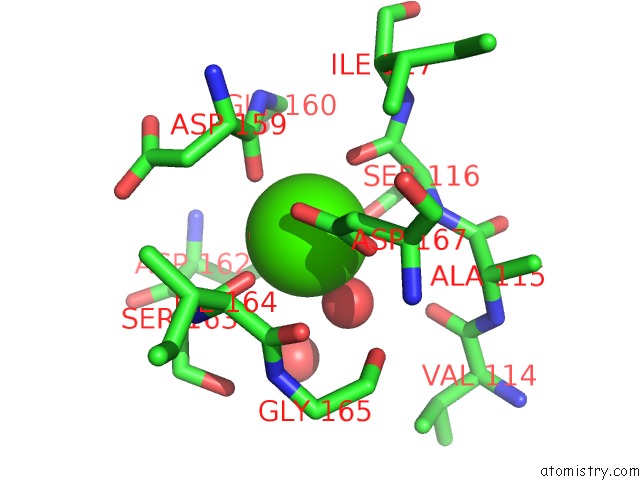
Mono view
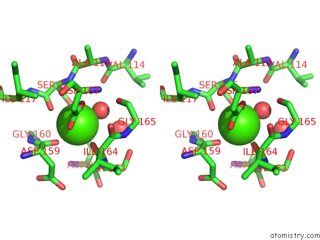
Stereo pair view
Mono view
Stereo pair view
A full contact list of Calcium with other atoms in the Ca binding
site number 1 of The Structure of CBM51 From Clostridium Perfringens GH95 in Complex with Methyl-Galactose within 5.0Å range:
|
Reference:
K.Gregg,
R.Finn,
D.W.Abbott,
A.B.Boraston.
Divergent Modes of Glycan Recognition By A New Family of Carbohydrate-Binding Modules J.Biol.Chem. V. 283 12604 2008.
ISSN: ISSN 0021-9258
PubMed: 18292090
DOI: 10.1074/JBC.M709865200
Page generated: Tue Jul 8 08:40:35 2025
ISSN: ISSN 0021-9258
PubMed: 18292090
DOI: 10.1074/JBC.M709865200
Last articles
Fe in 6CO5Fe in 6CL6
Fe in 6CL5
Fe in 6CKE
Fe in 6CIZ
Fe in 6CIR
Fe in 6CIQ
Fe in 6CDK
Fe in 6CII
Fe in 6CFW