Calcium »
PDB 2w3k-2whk »
2w9d »
Calcium in PDB 2w9d: Structure of Fab Fragment of the Icsm 18 - Anti-Prp Therapeutic Antibody at 1.57 A Resolution.
Protein crystallography data
The structure of Structure of Fab Fragment of the Icsm 18 - Anti-Prp Therapeutic Antibody at 1.57 A Resolution., PDB code: 2w9d
was solved by
S.V.Antonyuk,
C.R.Trevitt,
R.W.Strange,
G.S.Jackson,
D.Sangar,
M.Batchelor,
S.Jones,
T.Georgiou,
S.Cooper,
C.Fraser,
A.Khalili-Shirazi,
A.R.Clarke,
S.S.Hasnain,
J.Collinge,
with X-Ray Crystallography technique. A brief refinement statistics is given in the table below:
| Resolution Low / High (Å) | 27.00 / 1.57 |
| Space group | P 21 21 21 |
| Cell size a, b, c (Å), α, β, γ (°) | 36.784, 84.710, 127.852, 90.00, 90.00, 90.00 |
| R / Rfree (%) | 18.4 / 21.7 |
Calcium Binding Sites:
The binding sites of Calcium atom in the Structure of Fab Fragment of the Icsm 18 - Anti-Prp Therapeutic Antibody at 1.57 A Resolution.
(pdb code 2w9d). This binding sites where shown within
5.0 Angstroms radius around Calcium atom.
In total only one binding site of Calcium was determined in the Structure of Fab Fragment of the Icsm 18 - Anti-Prp Therapeutic Antibody at 1.57 A Resolution., PDB code: 2w9d:
In total only one binding site of Calcium was determined in the Structure of Fab Fragment of the Icsm 18 - Anti-Prp Therapeutic Antibody at 1.57 A Resolution., PDB code: 2w9d:
Calcium binding site 1 out of 1 in 2w9d
Go back to
Calcium binding site 1 out
of 1 in the Structure of Fab Fragment of the Icsm 18 - Anti-Prp Therapeutic Antibody at 1.57 A Resolution.
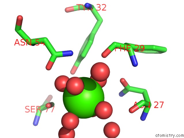
Mono view
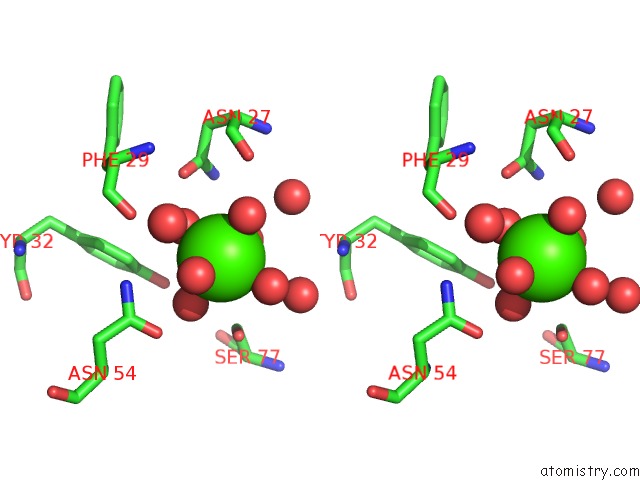
Stereo pair view
Mono view
Stereo pair view
A full contact list of Calcium with other atoms in the Ca binding
site number 1 of Structure of Fab Fragment of the Icsm 18 - Anti-Prp Therapeutic Antibody at 1.57 A Resolution. within 5.0Å range:
|
Reference:
S.V.Antonyuk,
C.R.Trevitt,
R.W.Strange,
G.S.Jackson,
D.Sangar,
M.Batchelor,
S.Cooper,
C.Fraser,
S.Jones,
T.Georgiou,
A.Khalili-Shirazi,
A.R.Clarke,
S.S.Hasnain,
J.Collinge.
Crystal Structure of Human Prion Protein Bound to A Therapeutic Antibody. Proc.Natl.Acad.Sci.Usa V. 106 2554 2009.
ISSN: ISSN 0027-8424
PubMed: 19204296
DOI: 10.1073/PNAS.0809170106
Page generated: Tue Jul 8 09:01:29 2025
ISSN: ISSN 0027-8424
PubMed: 19204296
DOI: 10.1073/PNAS.0809170106
Last articles
Mg in 4W5OMg in 4W5J
Mg in 4W5N
Mg in 4V2I
Mg in 4V3R
Mg in 4V26
Mg in 4V2G
Mg in 4V1T
Mg in 4V25
Mg in 4V1V